Nath Binita, Bidkar Anil P, Kumar Vikash, Dalal Amaresh, Jolly Mohit Kumar, Ghosh Siddhartha S, Biswas Gautam
Department of Mechanical Engineering, Indian Institute of Technology Guwahati, Guwahati 781 039, India.
Department of Biosciences and Bioengineering, Indian Institute of Technology Guwahati, Guwahati 781 039, India.
J Clin Med. 2019 Aug 9;8(8):1194. doi: 10.3390/jcm8081194.
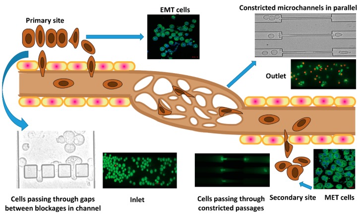
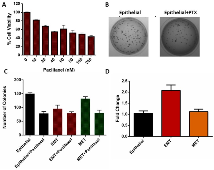
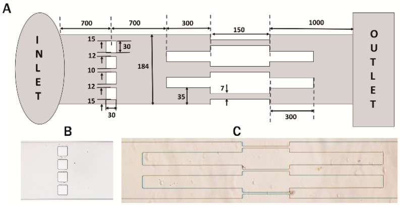
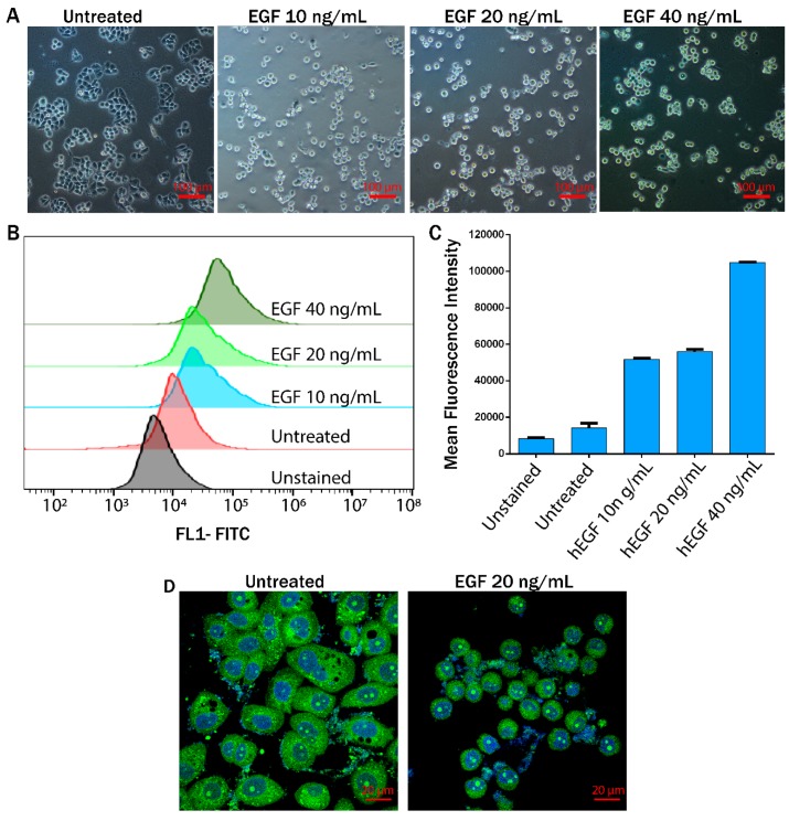
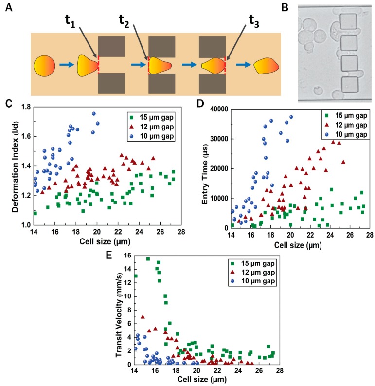
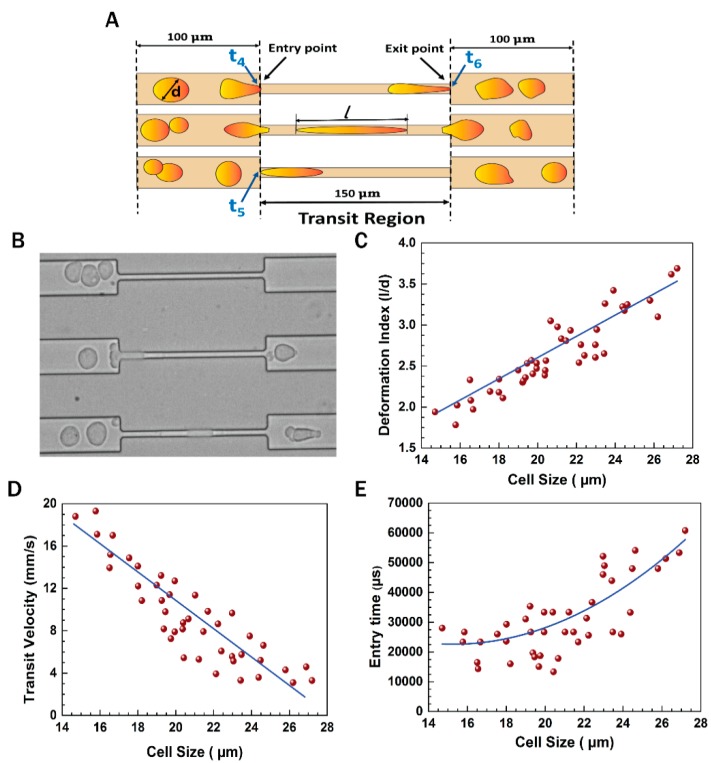
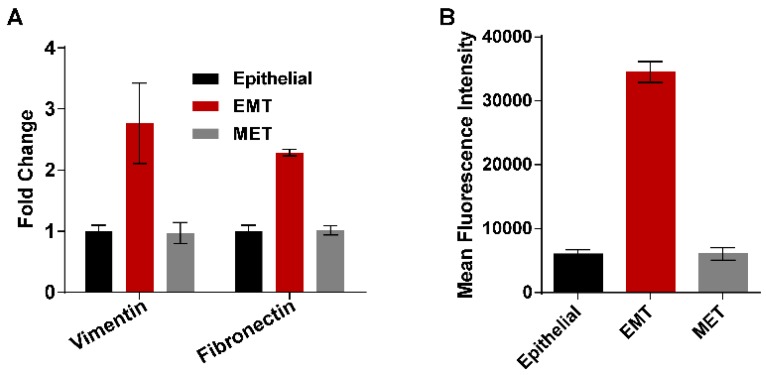
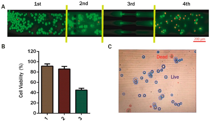
Epithelial to mesenchymal transition (EMT) induces cell migration, invasion, and drug resistance, and consequently, contributes to cancer metastasis and disease aggressiveness. This study attempted to address crucial biological parameters to correlate EMT and drug-treated cancer cells traversing through microcapillaries, reminiscent of metastatic conditions. MDA-MB-468 breast cancer cells induced to undergo EMT by treatment with 20 ng/mL of epidermal growth factor (EGF) were initially passed through several blockages and then through a constricted microchannel, mimicking the flow of invasive metastatic cells through constricted blood microcapillaries. EMT cells acquired enhanced migratory properties and retained 50% viability, even after migration through wells 10-15 μm in size and a constricted passage of 7 μm and 150 μm in length at a constant flow rate of 50 μL/h. The hydrodynamic properties revealed cellular deformation with a deformation index, average transit velocity, and entry time of 2.45, 12.3 mm/s, and 31,000 μs, respectively for a cell of average diameter 19 μm passing through one of the 7 μm constricted sections. Interestingly, cells collected at the channel outlet regained epithelial character, undergoing reverse transition (mesenchymal to epithelial transition, MET) in the absence of EGF. Remarkably, real-time polymerase chain reaction (PCR) analysis confirmed increases of 2- and 2.7-fold in the vimentin and fibronectin expression in EMT cells, respectively; however, their expression reduced to basal level in the MET cells. A scratch assay revealed the pronounced migratory nature of EMT cells compared with MET cells. Furthermore, the number of colonies formed from EMT cells and paclitaxel-treated EMT cells after passing through a constriction were found to be 95 ± 10 and 79 ± 4, respectively, confirming that the EMT cells were more drug resistant with a concomitant two-fold higher expression of the multi-drug resistance (MDR1) gene. Our results highlight the hydrodynamic and drug-evading properties of cells that have undergone an EMT, when passed through a constricted microcapillary that mimics their journey in blood circulation.
上皮-间质转化(EMT)会诱导细胞迁移、侵袭及产生耐药性,进而促使癌症转移和疾病进展。本研究试图探讨关键生物学参数,以关联EMT与经药物处理后穿越微毛细血管的癌细胞,这类似于转移状态。用20 ng/mL表皮生长因子(EGF)处理诱导发生EMT的MDA-MB-468乳腺癌细胞,首先使其通过若干阻塞物,然后通过一个狭窄微通道,模拟侵袭性转移细胞通过狭窄血液微毛细血管的流动。即使在以50 μL/h的恒定流速通过尺寸为10 - 15 μm的孔以及长度为7 μm和150 μm的狭窄通道后,EMT细胞仍获得了增强的迁移特性并保持50%的活力。流体动力学特性显示,对于平均直径为19 μm的细胞通过其中一个7 μm狭窄部分,其细胞变形指数、平均通过速度和进入时间分别为2.45、12.3 mm/s和31,000 μs。有趣的是,在通道出口收集的细胞在没有EGF的情况下恢复了上皮特征,发生了逆向转化(间质-上皮转化,MET)。值得注意的是,实时聚合酶链反应(PCR)分析证实,EMT细胞中波形蛋白和纤连蛋白的表达分别增加了2倍和2.7倍;然而,它们在MET细胞中的表达降至基础水平。划痕试验显示,与MET细胞相比,EMT细胞具有明显的迁移特性。此外,发现通过狭窄通道后,EMT细胞和经紫杉醇处理的EMT细胞形成的集落数分别为95±10和79±4,证实EMT细胞具有更强的耐药性,同时多药耐药(MDR1)基因的表达高出两倍。我们的结果突出了经历EMT的细胞在通过模拟其血液循环旅程的狭窄微毛细血管时的流体动力学和逃避药物的特性。