School of Molecular Biosciences, Washington State University, Pullman, WA, United States of America.
Center for Reproductive Biology, Washington State University, Pullman, WA, United States of America.
PLoS Genet. 2018 Nov 28;14(11):e1007823. doi: 10.1371/journal.pgen.1007823. eCollection 2018 Nov.
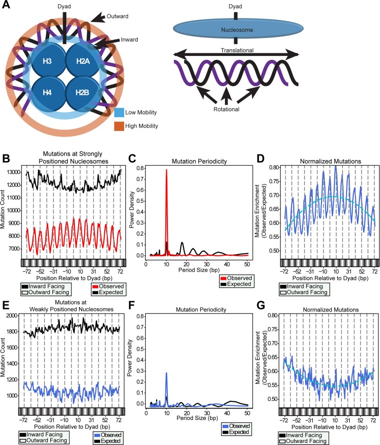
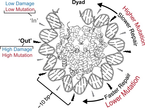
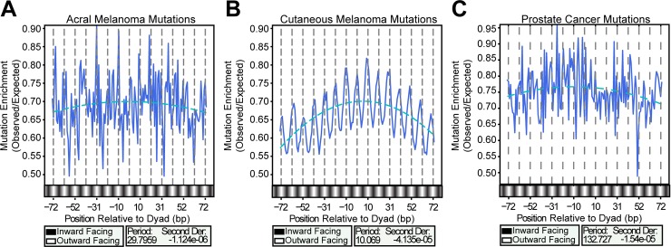
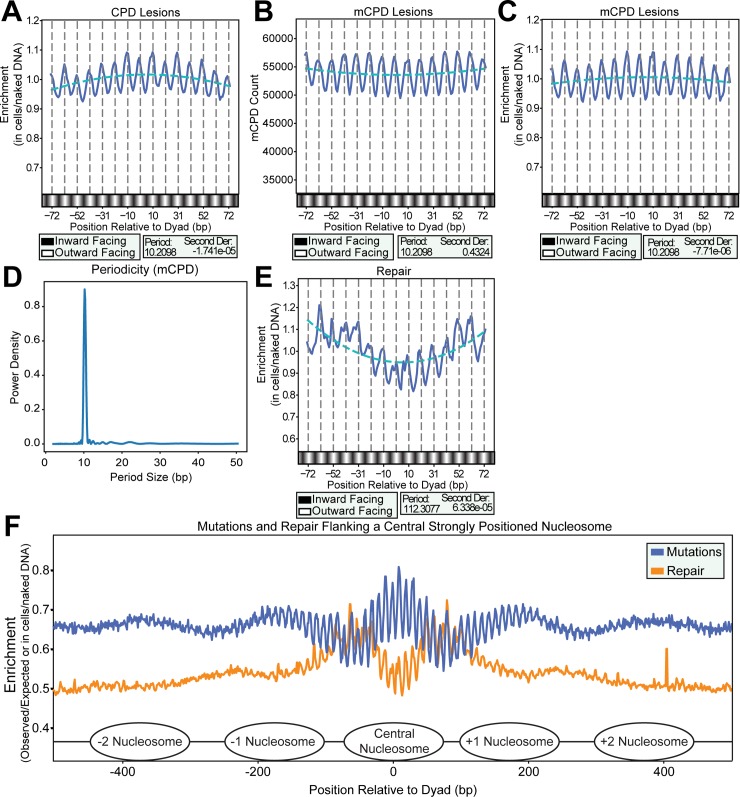
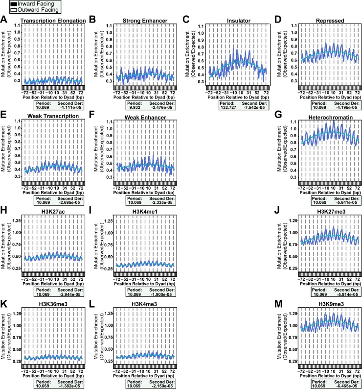
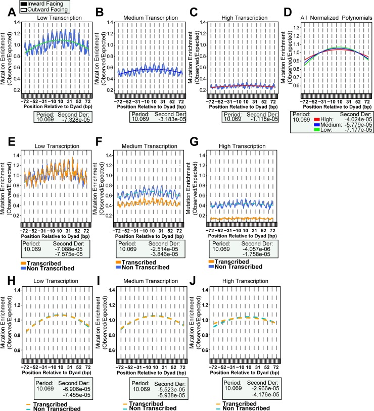
Ultraviolet (UV) light-induced mutations are unevenly distributed across skin cancer genomes, but the molecular mechanisms responsible for this heterogeneity are not fully understood. Here, we assessed how nucleosome structure impacts the positions of UV-induced mutations in human melanomas. Analysis of mutation positions from cutaneous melanomas within strongly positioned nucleosomes revealed a striking ~10 base pair (bp) oscillation in mutation density with peaks occurring at dinucleotides facing away from the histone octamer. Additionally, higher mutation density at the nucleosome dyad generated an overarching "translational curvature" across the 147 bp of DNA that constitutes the nucleosome core particle. This periodicity and curvature cannot be explained by sequence biases in nucleosomal DNA. Instead, our genome-wide map of UV-induced cyclobutane pyrimidine dimers (CPDs) indicates that CPD formation is elevated at outward facing dinucleotides, mirroring the oscillation of mutation density within nucleosome-bound DNA. Nucleotide excision repair (NER) activity, as measured by XR-seq, inversely correlated with the curvature of mutation density associated with the translational setting of the nucleosome. While the 10 bp periodicity of mutations is maintained across nucleosomes regardless of chromatin state, histone modifications, and transcription levels, overall mutation density and curvature across the core particle increased with lower transcription levels. Our observations suggest structural conformations of DNA promote CPD formation at specific sites within nucleosomes, and steric hindrance progressively limits lesion repair towards the nucleosome dyad. Both mechanisms create a unique extended mutation signature within strongly positioned nucleosomes across the human genome.
紫外线(UV)诱导的突变在皮肤癌基因组中分布不均匀,但导致这种异质性的分子机制尚不完全清楚。在这里,我们评估了核小体结构如何影响人类黑色素瘤中 UV 诱导突变的位置。对来自皮肤黑色素瘤的突变位置的分析表明,在强烈定位的核小体中,突变密度存在明显的~10 个碱基对(bp)的摆动,突变峰出现在远离组蛋白八聚体的二核苷酸上。此外,核小体二分体处更高的突变密度产生了跨越构成核小体核心颗粒的 147bp DNA 的总体“平移曲率”。这种周期性和曲率不能用核小体 DNA 中的序列偏倚来解释。相反,我们对 UV 诱导的环丁烷嘧啶二聚体(CPD)的全基因组图谱表明,CPD 的形成在面向外侧的二核苷酸处升高,与核小体结合 DNA 内突变密度的摆动相吻合。核苷酸切除修复(NER)活性,如 XR-seq 测量所示,与与核小体翻译设置相关的突变密度的曲率呈负相关。虽然突变的 10bp 周期性在无论染色质状态、组蛋白修饰和转录水平如何都在核小体中保持不变,但核心颗粒中突变密度和曲率的整体增加与转录水平降低有关。我们的观察结果表明,DNA 的结构构象促进了核小体中特定部位的 CPD 形成,并且随着向核小体二分体的推进,空间位阻逐渐限制了损伤修复。这两种机制在整个人类基因组中为强烈定位的核小体创造了独特的扩展突变特征。