Romero-Molina Carmen, Navarro Victoria, Sanchez-Varo Raquel, Jimenez Sebastian, Fernandez-Valenzuela Juan J, Sanchez-Mico Maria V, Muñoz-Castro Clara, Gutierrez Antonia, Vitorica Javier, Vizuete Marisa
Departamento Bioquimica y Biologia Molecular, Facultad de Farmacia, Universidad de Sevilla, Seville, Spain.
Instituto de Biomedicina de Sevilla, Hospital Universitario Virgen del Rocio, CSIC, Universidad de Sevilla, Seville, Spain.
Front Cell Neurosci. 2018 Nov 14;12:421. doi: 10.3389/fncel.2018.00421. eCollection 2018.
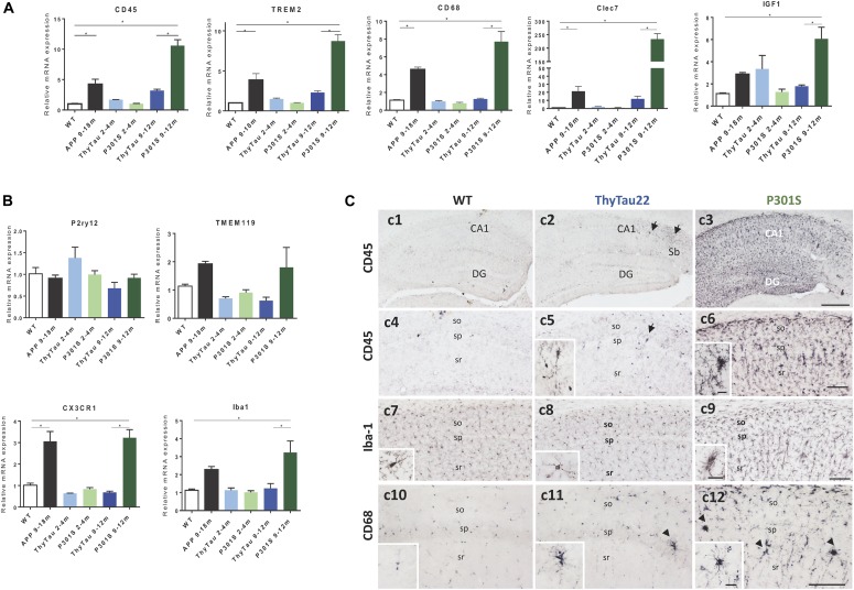
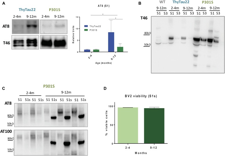
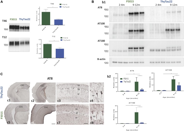
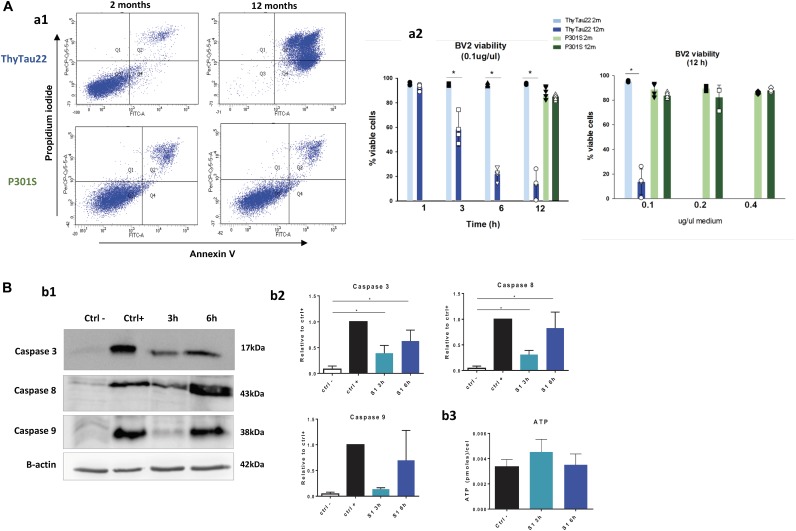
Microglial cells are crucial players in the pathological process of neurodegenerative diseases, such as Alzheimer's disease (AD). Microglial response in AD has been principally studied in relation to amyloid-beta pathology but, comparatively, little is known about inflammatory processes associated to tau pathology. In the hippocampus of AD patients, where tau pathology is more prominent than amyloid-beta pathology, a microglial degenerative process has been reported. In this work, we have directly compared the microglial response in two different transgenic tau mouse models: ThyTau22 and P301S. Surprisingly, these two models showed important differences in the microglial profile and tau pathology. Where ThyTau22 hippocampus manifested mild microglial activation, P301S mice exhibited a strong microglial response in parallel with high phospho-tau accumulation. This differential phospho-tau expression could account for the different microglial response in these two tau strains. However, soluble (S1) fractions from ThyTau22 hippocampus presented relatively high content of soluble phospho-tau (AT8-positive) and were highly toxic for microglial cells , whereas the correspondent S1 fractions from P301S mice displayed low soluble phospho-tau levels and were not toxic for microglial cells. Therefore, not only the expression levels but the aggregation of phospho-tau should differ between both models. In fact, most of tau forms in the P301S mice were aggregated and, in consequence, forming insoluble tau species. We conclude that different factors as tau mutations, accumulation, phosphorylation, and/or aggregation could account for the distinct microglial responses observed in these two tau models. For this reason, deciphering the molecular nature of toxic tau species for microglial cells might be a promising therapeutic approach in order to restore the deficient immunological protection observed in AD hippocampus.
小胶质细胞是神经退行性疾病(如阿尔茨海默病,AD)病理过程中的关键参与者。AD中的小胶质细胞反应主要是针对β-淀粉样蛋白病理进行研究的,但相比之下,关于与tau病理相关的炎症过程知之甚少。在AD患者的海马体中,tau病理比β-淀粉样蛋白病理更为突出,已有报道称存在小胶质细胞变性过程。在这项研究中,我们直接比较了两种不同的转基因tau小鼠模型(ThyTau22和P301S)中的小胶质细胞反应。令人惊讶的是,这两种模型在小胶质细胞特征和tau病理方面表现出重要差异。ThyTau22海马体表现出轻度的小胶质细胞激活,而P301S小鼠则表现出强烈的小胶质细胞反应,同时伴有高磷酸化tau的积累。这种磷酸化tau表达的差异可能解释了这两种tau菌株中小胶质细胞反应的不同。然而,ThyTau22海马体的可溶性(S1)组分呈现出相对较高的可溶性磷酸化tau(AT8阳性)含量,并且对小胶质细胞具有高度毒性,而P301S小鼠相应的S1组分显示出低可溶性磷酸化tau水平,对小胶质细胞无毒。因此,不仅两种模型中磷酸化tau的表达水平不同,其聚集情况也应有所差异。事实上,P301S小鼠中的大多数tau形式都发生了聚集,因此形成了不溶性tau物种。我们得出结论,tau突变、积累、磷酸化和/或聚集等不同因素可能解释了在这两种tau模型中观察到的不同小胶质细胞反应。因此,解读对小胶质细胞有毒性的tau物种的分子性质可能是一种有前景的治疗方法,以便恢复在AD海马体中观察到的免疫保护缺陷。