• 文献检索
  • 文档翻译
  • 深度研究
  • 学术资讯
  • Suppr Zotero 插件Zotero 插件
  • 邀请有礼
  • 套餐&价格
  • 历史记录
应用&插件
Suppr Zotero 插件Zotero 插件浏览器插件Mac 客户端Windows 客户端微信小程序
定价
高级版会员购买积分包购买API积分包
服务
文献检索文档翻译深度研究API 文档MCP 服务
关于我们
关于 Suppr公司介绍联系我们用户协议隐私条款
关注我们

Suppr 超能文献

核心技术专利:CN118964589B侵权必究
粤ICP备2023148730 号-1Suppr @ 2026

文献检索

告别复杂PubMed语法,用中文像聊天一样搜索,搜遍4000万医学文献。AI智能推荐,让科研检索更轻松。

立即免费搜索

文件翻译

保留排版,准确专业,支持PDF/Word/PPT等文件格式,支持 12+语言互译。

免费翻译文档

深度研究

AI帮你快速写综述,25分钟生成高质量综述,智能提取关键信息,辅助科研写作。

立即免费体验

心血管 MRI 评估主动脉瓣狭窄的主动脉僵硬度:二叶瓣和三叶瓣的比较。

Aortic stiffness in aortic stenosis assessed by cardiovascular MRI: a comparison between bicuspid and tricuspid valves.

机构信息

Department of Cardiovascular Sciences, University of Leicester and Cardiovascular Theme, NIHR Leicester Biomedical Research Centre, Glenfield Hospital, Groby road, Leicester, LE3 9QP, UK.

Xinapse Systems Ltd., West Bergholt, Essex, CO6 3BW, UK.

出版信息

Eur Radiol. 2019 May;29(5):2340-2349. doi: 10.1007/s00330-018-5775-6. Epub 2018 Nov 28.

DOI:10.1007/s00330-018-5775-6
PMID:30488106
原文链接:https://pmc.ncbi.nlm.nih.gov/articles/PMC6443917/
Abstract

OBJECTIVES

To compare aortic size and stiffness parameters on MRI between bicuspid aortic valve (BAV) and tricuspid aortic valve (TAV) patients with aortic stenosis (AS).

METHODS

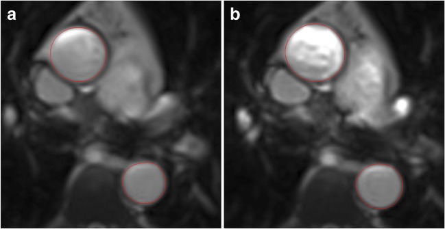
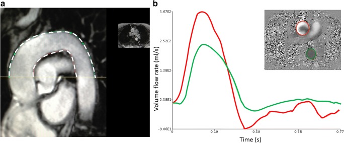
MRI was performed in 174 patients with asymptomatic moderate-severe AS (mean AVAI 0.57 ± 0.14 cm/m) and 23 controls on 3T scanners. Valve morphology was available/analysable in 169 patients: 63 BAV (41 type-I, 22 type-II) and 106 TAV. Aortic cross-sectional areas were measured at the level of the pulmonary artery bifurcation. The ascending and descending aorta (AA, DA) distensibility, and pulse wave velocity (PWV) around the aortic arch were calculated.

RESULTS

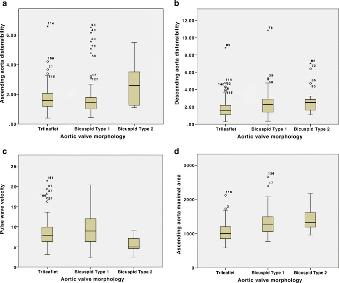
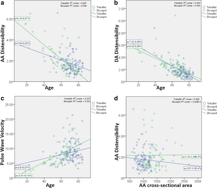
The AA and DA areas were lower in the controls, with no difference in DA distensibility or PWV, but slightly lower AA distensibility than in the patient group. With increasing age, there was a decrease in distensibility and an increase in PWV. After correcting for age, the AA maximum cross-sectional area was higher in bicuspid vs. tricuspid patients (12.97 [11.10, 15.59] vs. 10.06 [8.57, 12.04] cm, p < 0.001), but there were no significant differences in AA distensibility (p = 0.099), DA distensibility (p = 0.498) or PWV (p = 0.235). Patients with BAV type-II valves demonstrated a significantly higher AA distensibility and lower PWV compared to type-I, despite a trend towards higher AA area.

CONCLUSIONS

In patients with significant AS, BAV patients do not have increased aortic stiffness compared to those with TAV despite increased ascending aortic dimensions. Those with type-II BAV have less aortic stiffness despite greater dimensions. These results demonstrate a dissociation between aortic dilatation and stiffness and suggest that altered flow patterns may play a role.

KEY POINTS

• Both cellular abnormalities secondary to genetic differences and abnormal flow patterns have been implicated in the pathophysiology of aortic dilatation and increased vascular complications associated with bicuspid aortic valves (BAV). • We demonstrate an increased ascending aortic size in patients with BAV and moderate to severe AS compared to TAV and controls, but no difference in aortic stiffness parameters, therefore suggesting a dissociation between dilatation and stiffness. • Sub-group analysis showed greater aortic size but lower stiffness parameters in those with BAV type-II AS compared to BAV type-I.

摘要

目的

比较 MRI 显示的二叶式主动脉瓣(BAV)和三叶式主动脉瓣(TAV)主动脉瓣狭窄(AS)患者的主动脉大小和僵硬度参数。

方法

在 3T 扫描仪上对 174 例无症状中重度 AS(平均 AVAI 0.57±0.14 cm/m)患者和 23 例对照者进行 MRI 检查。在 169 例患者中可获得/可分析瓣膜形态:63 例 BAV(41 例 I 型,22 例 II 型)和 106 例 TAV。在肺动脉分叉处测量主动脉的横截面积。计算升主动脉(AA)和降主动脉(DA)的可扩展性以及主动脉弓周围的脉搏波速度(PWV)。

结果

对照组的 AA 和 DA 面积较低,DA 扩展性或 PWV 无差异,但 AA 扩展性略低于患者组。随着年龄的增长,可扩展性降低,PWV 增加。校正年龄后,二叶瓣患者的 AA 最大横截面积高于三叶瓣患者(12.97[11.10,15.59] vs. 10.06[8.57,12.04] cm,p<0.001),但 AA 扩展性(p=0.099)、DA 扩展性(p=0.498)或 PWV(p=0.235)无显著差异。与 I 型相比,II 型 BAV 患者的 AA 扩展性更高,PWV 更低,尽管 AA 面积有增大趋势。

结论

在有明显 AS 的患者中,与 TAV 患者相比,BAV 患者的主动脉僵硬度没有增加,尽管升主动脉尺寸增大。尽管二叶瓣患者的升主动脉尺寸较大,但主动脉僵硬度较低。这些结果表明主动脉扩张和僵硬度之间存在分离,并提示血流模式的改变可能起作用。

关键点

  1. 细胞异常继发于遗传差异和异常的血流模式,与二叶式主动脉瓣(BAV)相关的主动脉扩张和增加的血管并发症的病理生理学有关。

  2. 我们发现,与 TAV 和对照组相比,二叶瓣主动脉瓣狭窄(AS)患者的升主动脉尺寸增大,但主动脉僵硬度参数无差异,因此提示扩张和僵硬度之间存在分离。

  3. 亚组分析显示,与二叶瓣 I 型相比,二叶瓣 II 型 AS 患者的主动脉尺寸更大,但僵硬度参数更低。

https://cdn.ncbi.nlm.nih.gov/pmc/blobs/00b1/6443917/51ea37bd0858/330_2018_5775_Fig4_HTML.jpg
https://cdn.ncbi.nlm.nih.gov/pmc/blobs/00b1/6443917/9492b0dc55a9/330_2018_5775_Fig1_HTML.jpg
https://cdn.ncbi.nlm.nih.gov/pmc/blobs/00b1/6443917/4a0493708611/330_2018_5775_Fig2_HTML.jpg
https://cdn.ncbi.nlm.nih.gov/pmc/blobs/00b1/6443917/94d86a9eb651/330_2018_5775_Fig3_HTML.jpg
https://cdn.ncbi.nlm.nih.gov/pmc/blobs/00b1/6443917/51ea37bd0858/330_2018_5775_Fig4_HTML.jpg

相似文献

1
Aortic stiffness in aortic stenosis assessed by cardiovascular MRI: a comparison between bicuspid and tricuspid valves.心血管 MRI 评估主动脉瓣狭窄的主动脉僵硬度:二叶瓣和三叶瓣的比较。
Eur Radiol. 2019 May;29(5):2340-2349. doi: 10.1007/s00330-018-5775-6. Epub 2018 Nov 28.
2
Influence of Aortic Dilation on the Regional Aortic Stiffness of Bicuspid Aortic Valve Assessed by 4-Dimensional Flow Cardiac Magnetic Resonance: Comparison With Marfan Syndrome and Degenerative Aortic Aneurysm.4D 血流心脏磁共振评估主动脉扩张对二叶式主动脉瓣区域性主动脉僵硬度的影响:与马凡综合征和退行性主动脉瘤的比较。
JACC Cardiovasc Imaging. 2019 Jun;12(6):1020-1029. doi: 10.1016/j.jcmg.2018.03.017. Epub 2018 May 16.
3
Comprehensive assessment of local and regional aortic stiffness in patients with tricuspid or bicuspid aortic valve aortopathy using magnetic resonance imaging.应用磁共振成像技术全面评估三尖瓣或二叶式主动脉瓣主动脉瓣病变患者的局部和区域性主动脉僵硬度。
Int J Cardiol. 2021 Mar 1;326:206-212. doi: 10.1016/j.ijcard.2020.11.046. Epub 2020 Nov 28.
4
Patterns of ascending aortic dilatation and predictors of surgical replacement of the aorta: A comparison of bicuspid and tricuspid aortic valve patients over eight years of follow-up.升主动脉扩张模式和主动脉手术置换的预测因素:在八年的随访中,对二叶式主动脉瓣和三叶式主动脉瓣患者的比较。
J Mol Cell Cardiol. 2019 Oct;135:31-39. doi: 10.1016/j.yjmcc.2019.07.010. Epub 2019 Jul 23.
5
Proximal aortic stiffening in Turner patients may be present before dilation can be detected: a segmental functional MRI study.特纳综合征患者的主动脉近端僵硬可能在检测到扩张之前就已存在:一项分段功能磁共振成像研究。
J Cardiovasc Magn Reson. 2017 Feb 13;19(1):27. doi: 10.1186/s12968-017-0331-0.
6
Aortic stiffness in adolescent Turner and Marfan syndrome patients.青少年特纳综合征和马凡综合征患者的主动脉僵硬度。
Eur J Cardiothorac Surg. 2018 Nov 1;54(5):926-932. doi: 10.1093/ejcts/ezy168.
7
Aortic wall thickness in dilated ascending aorta: Comparison between tricuspid and bicuspid aortic valve.扩张性升主动脉的主动脉壁厚度:三尖瓣主动脉瓣与二瓣化主动脉瓣的比较。
Arch Cardiovasc Dis. 2023 Nov;116(11):498-505. doi: 10.1016/j.acvd.2023.08.003. Epub 2023 Sep 14.
8
Aortic dimensions in relation to bicuspid and tricuspid aortic valve pathology.与二叶式和三叶式主动脉瓣病变相关的主动脉尺寸
J Heart Valve Dis. 2014 Jul;23(4):463-72.
9
Dilatation of the ascending aorta in bicuspid aortic valve disease: a magnetic resonance imaging study.二叶式主动脉瓣疾病中升主动脉扩张:一项磁共振成像研究
Clin Res Cardiol. 2009 Feb;98(2):114-20. doi: 10.1007/s00392-008-0731-0. Epub 2008 Dec 12.
10
Augmentation index and aortic stiffness in bicuspid aortic valve patients with non-dilated proximal aortas.二叶式主动脉瓣患者非扩张性升主动脉的增强指数和主动脉僵硬度。
BMC Cardiovasc Disord. 2013 Mar 15;13:19. doi: 10.1186/1471-2261-13-19.

引用本文的文献

1
Resource efficient aortic distensibility calculation by end to end spatiotemporal learning of aortic lumen from multicentre multivendor multidisease CMR images.基于端到端时空学习的多中心多供应商多病种 CMR 图像主动脉管腔的资源高效主动脉可扩张性计算。
Sci Rep. 2023 Dec 8;13(1):21794. doi: 10.1038/s41598-023-48986-6.
2
Feasibility of Wave Intensity Analysis from 4D Cardiovascular Magnetic Resonance Imaging Data.基于四维心血管磁共振成像数据的波强度分析的可行性
Bioengineering (Basel). 2023 May 31;10(6):662. doi: 10.3390/bioengineering10060662.
3
Aortic wall shear stress in bicuspid aortic valve disease-10-year follow-up.

本文引用的文献

1
Comparison of exercise testing and CMR measured myocardial perfusion reserve for predicting outcome in asymptomatic aortic stenosis: the PRognostic Importance of MIcrovascular Dysfunction in Aortic Stenosis (PRIMID AS) Study.运动试验与心脏磁共振成像测量心肌灌注储备对预测无症状性主动脉瓣狭窄预后的比较:主动脉瓣狭窄微血管功能障碍的预后重要性(PRIMID AS)研究
Eur Heart J. 2017 Apr 21;38(16):1222-1229. doi: 10.1093/eurheartj/ehx001.
2
Myocardial T1 and extracellular volume fraction measurement in asymptomatic patients with aortic stenosis: reproducibility and comparison with age-matched controls.无症状主动脉瓣狭窄患者的心肌T1和细胞外容积分数测量:可重复性及与年龄匹配对照组的比较
Eur Heart J Cardiovasc Imaging. 2015 Jul;16(7):763-70. doi: 10.1093/ehjci/jev007. Epub 2015 Feb 13.
3
二叶式主动脉瓣疾病中主动脉壁剪切应力的10年随访
Cardiovasc Diagn Ther. 2023 Feb 28;13(1):38-50. doi: 10.21037/cdt-22-477. Epub 2023 Feb 10.
4
Clinical implications of the biomechanics of bicuspid aortic valve and bicuspid aortopathy.二叶式主动脉瓣及二叶式主动脉病变生物力学的临床意义
Front Cardiovasc Med. 2022 Aug 12;9:922353. doi: 10.3389/fcvm.2022.922353. eCollection 2022.
5
The role of 4-dimensional flow in the assessment of bicuspid aortic valve and its valvulo-aortopathies.四维血流在二叶式主动脉瓣及其瓣-主动脉病变评估中的作用。
Br J Radiol. 2022 Oct 1;95(1139):20220123. doi: 10.1259/bjr.20220123. Epub 2022 Aug 3.
6
PHACTR1 modulates vascular compliance but not endothelial function: a translational study.PHACTR1 调节血管顺应性但不影响血管内皮功能:一项转化研究。
Cardiovasc Res. 2023 Mar 31;119(2):599-610. doi: 10.1093/cvr/cvac092.
7
Global Aortic Pulse Wave Velocity is Unchanged in Bicuspid Aortopathy With Normal Valve Function but Elevated in Patients With Aortic Valve Stenosis: Insights From a 4D Flow MRI Study of 597 Subjects.全球主动脉脉搏波速度在瓣叶功能正常的二叶式主动脉瓣病变中保持不变,但在主动脉瓣狭窄患者中升高:来自 597 例受试者的 4D 血流 MRI 研究的结果。
J Magn Reson Imaging. 2023 Jan;57(1):126-136. doi: 10.1002/jmri.28266. Epub 2022 May 28.
8
Arterial Stiffness in Aortic Stenosis.主动脉瓣狭窄中的动脉僵硬度
Arq Bras Cardiol. 2021 Mar;116(3):483-484. doi: 10.36660/abc.20201234.
9
Evaluation of Plaque Characteristics and Inflammation Using Magnetic Resonance Imaging.使用磁共振成像评估斑块特征和炎症
Biomedicines. 2021 Feb 12;9(2):185. doi: 10.3390/biomedicines9020185.
Subclinical diastolic dysfunction in young adults with Type 2 diabetes mellitus: a multiparametric contrast-enhanced cardiovascular magnetic resonance pilot study assessing potential mechanisms.2型糖尿病青年成人的亚临床舒张功能障碍:一项多参数对比增强心血管磁共振初步研究,评估潜在机制
Eur Heart J Cardiovasc Imaging. 2014 Nov;15(11):1263-9. doi: 10.1093/ehjci/jeu121. Epub 2014 Jun 26.
4
Rationale and design of the PRognostic Importance of MIcrovascular Dysfunction in asymptomatic patients with Aortic Stenosis (PRIMID-AS): a multicentre observational study with blinded investigations.无症状主动脉瓣狭窄患者微血管功能障碍预后重要性的研究(PRIMID-AS)的原理和设计:一项多中心观察性研究,具有盲法研究。
BMJ Open. 2013 Dec 18;3(12):e004348. doi: 10.1136/bmjopen-2013-004348.
5
Normal values of aortic dimensions, distensibility, and pulse wave velocity in children and young adults: a cross-sectional study.儿童和青年成年人的主动脉尺寸、可扩张性和脉搏波速度的正常值:一项横断面研究。
J Cardiovasc Magn Reson. 2012 Nov 14;14(1):77. doi: 10.1186/1532-429X-14-77.
6
Assessment of abdominal aortic aneurysm wall distensibility with electrocardiography-gated computed tomography.利用心电图门控计算机断层扫描评估腹主动脉瘤壁的扩张性
Ann Vasc Surg. 2011 Nov;25(8):1036-42. doi: 10.1016/j.avsg.2011.05.034.
7
Measuring aortic pulse wave velocity using high-field cardiovascular magnetic resonance: comparison of techniques.使用高场心血管磁共振测量主动脉脉搏波速度:技术比较。
J Cardiovasc Magn Reson. 2010 May 11;12(1):26. doi: 10.1186/1532-429X-12-26.
8
Bicuspid aortic valve: four-dimensional MR evaluation of ascending aortic systolic flow patterns.二叶式主动脉瓣:升主动脉收缩期血流模式的四维磁共振评估。
Radiology. 2010 Apr;255(1):53-61. doi: 10.1148/radiol.09091437.
9
Predictors of exercise capacity and symptoms in severe aortic stenosis.重度主动脉瓣狭窄患者运动能力和症状的预测因素
Eur J Echocardiogr. 2010 Jul;11(6):482-7. doi: 10.1093/ejechocard/jeq002. Epub 2010 Feb 6.
10
Hemodynamic predictors of aortic dilatation in bicuspid aortic valve by velocity-encoded cardiovascular magnetic resonance.速度编码心血管磁共振对二叶式主动脉瓣主动脉扩张的血流动力学预测因素。
J Cardiovasc Magn Reson. 2010 Jan 13;12(1):4. doi: 10.1186/1532-429X-12-4.