Department of Physics and Astronomy, Iowa State University, Ames, Iowa.
Molecular, Cellular, and Developmental Biology Interdepartmental Program, Molecular Biology Building, Ames, Iowa.
J Biophotonics. 2019 May;12(5):e201800351. doi: 10.1002/jbio.201800351. Epub 2019 Jan 28.
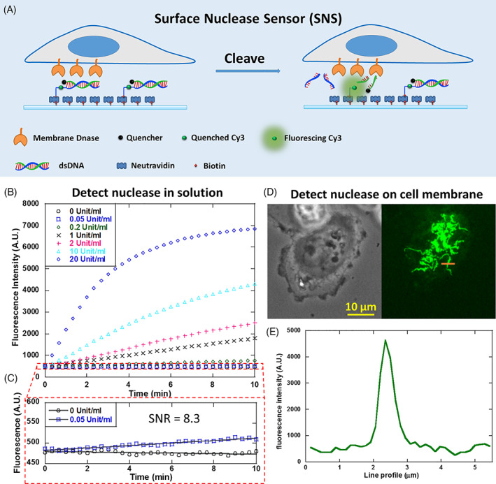
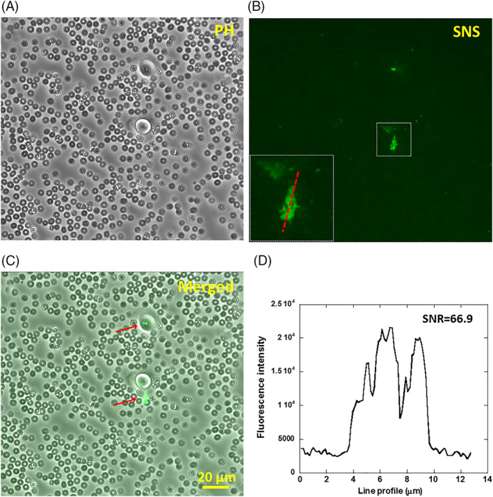
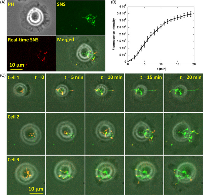
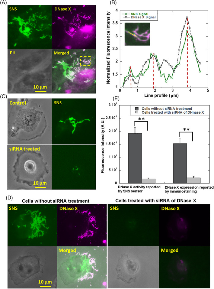
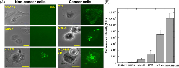
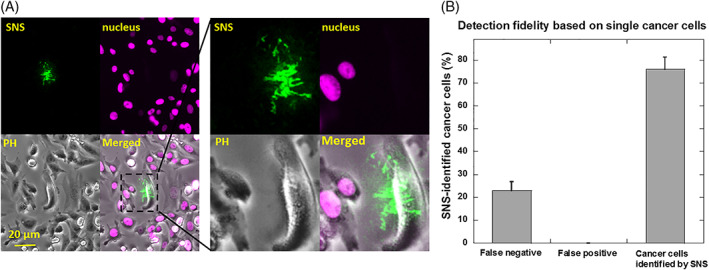
Nucleases are important enzymes that cleave nucleic acids and play critical roles in DNA repair, immune defense and potentially in cancer invasion. However, their spatial dynamics at subcellular level is much less studied. Here, we developed a surface-tethered nuclease sensor (SNS) which directly converts membrane-bound nuclease (MN) activity to fluorescent signal, therefore, mapping MN activity on cell adhesion sites with high resolution and sensitivity. With SNS, we studied MN activity on the ventral membrane of cancer cells, where MN activity initially occurs in punctate regions and advances in a coral-shaped pattern. In six tested cell-lines, the MN activity levels in cancer cells are significantly higher than those in non-cancer cells. We then tested SNS as a sensitive approach to detect cancer cells at single cell level. Single breast cancer cells were successfully detected from thousands of adherent non-cancer cells and from millions of non-adherent blood cells.
核酸酶是重要的酶类,能够切割核酸,在 DNA 修复、免疫防御等方面发挥关键作用,并且可能与癌症侵袭有关。然而,其在亚细胞水平的空间动力学变化研究还较少。在这里,我们开发了一种基于表面固定核酸酶传感器(SNS),它可以直接将膜结合核酸酶(MN)的活性转化为荧光信号,从而以高分辨率和灵敏度在细胞黏附部位绘制 MN 活性图。通过 SNS,我们研究了癌细胞腹膜上的 MN 活性,MN 活性最初出现在点状区域,并呈珊瑚状模式推进。在六种测试的细胞系中,癌细胞中的 MN 活性水平明显高于非癌细胞。然后,我们测试了 SNS 作为一种在单细胞水平检测癌细胞的敏感方法。成功地从数千个黏附的非癌细胞和数百万个非黏附的血细胞中检测到单个乳腺癌细胞。