Department of Radiology, Affiliated Southwest Hospital, Army Medical University, #29 Gaotanyan Main Street, Chongqing 400038, China.
Department of Psychology, Affiliated Southwest Hospital, Army Medical University, Chongqing, China.
Neuroimage Clin. 2019;21:101614. doi: 10.1016/j.nicl.2018.101614. Epub 2018 Nov 28.
Numerous cognitive and emotional functions are executed asymmetrically between the left and right hemispheres. Right hemisphere hyperactivity/left hemisphere hypoactivity often appears to be a feature in neuroimaging studies of depression. However, few studies have evaluated abnormalities in structural asymmetry in untreated patients with major depressive disorder (MDD).
In this study, 3-dimensional high-resolution structural magnetic resonance images were acquired from 35 treatment-naïve patients with MDD (mean age = 28.9 years, 22 females) and 35 normal controls. The asymmetry index in cortical thickness and subcortical volume were calculated based on an automated surface-based technique.
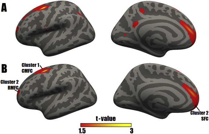
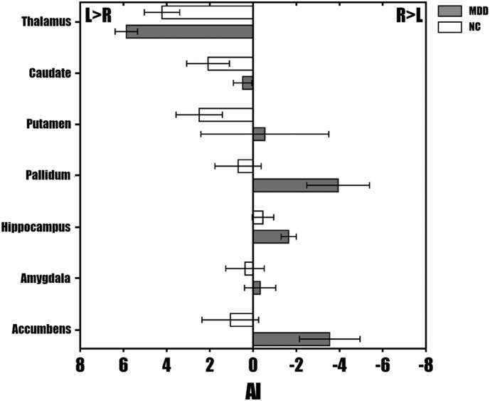
Abnormalities in structural asymmetry in patients with MDD were mainly located in the cortical-striatal-pallidal-thalamic circuit, including the superior frontal cortex, rostral middle frontal cortex, caudal middle frontal cortex, nucleus accumbens, pallidum and thalamus. No significant correlation was observed between symptom severity and asymmetric measurements.
These findings provide further evidence for the altered morphological interhemispheric imbalances in depression and these alterations were independent of depressive symptom severity, suggesting that cerebral asymmetry could be an appropriate indicator of morphological variations in mental disease.
许多认知和情感功能在大脑的左、右半球之间存在不对称性。右半球活动过度/左半球活动不足,这种现象在抑郁症的神经影像学研究中经常出现。然而,很少有研究评估未经治疗的重度抑郁症(MDD)患者结构不对称性的异常。
本研究对 35 名未经治疗的 MDD 患者(平均年龄 28.9 岁,22 名女性)和 35 名正常对照者进行了三维高分辨率结构磁共振成像扫描。基于自动表面技术计算皮质厚度和皮质下体积的不对称指数。
MDD 患者的结构不对称性异常主要位于皮质-纹状体-苍白球-丘脑回路,包括额上回、额中回前部、额中回后部、伏隔核、苍白球和丘脑。症状严重程度与不对称测量值之间无显著相关性。
这些发现为抑郁症中形态学半球间平衡改变提供了进一步的证据,这些改变与抑郁症状严重程度无关,表明大脑不对称性可能是精神疾病形态变化的一个合适指标。