• 文献检索
  • 文档翻译
  • 深度研究
  • 学术资讯
  • Suppr Zotero 插件Zotero 插件
  • 邀请有礼
  • 套餐&价格
  • 历史记录
应用&插件
Suppr Zotero 插件Zotero 插件浏览器插件Mac 客户端Windows 客户端微信小程序
定价
高级版会员购买积分包购买API积分包
服务
文献检索文档翻译深度研究API 文档MCP 服务
关于我们
关于 Suppr公司介绍联系我们用户协议隐私条款
关注我们

Suppr 超能文献

核心技术专利:CN118964589B侵权必究
粤ICP备2023148730 号-1Suppr @ 2026

文献检索

告别复杂PubMed语法,用中文像聊天一样搜索,搜遍4000万医学文献。AI智能推荐,让科研检索更轻松。

立即免费搜索

文件翻译

保留排版,准确专业,支持PDF/Word/PPT等文件格式,支持 12+语言互译。

免费翻译文档

深度研究

AI帮你快速写综述,25分钟生成高质量综述,智能提取关键信息,辅助科研写作。

立即免费体验

生物 corona 形成导致银纳米颗粒诱导的内质网应激。

Biocorona formation contributes to silver nanoparticle induced endoplasmic reticulum stress.

机构信息

Department of Pharmaceutical Sciences, Skaggs School of Pharmacy and Pharmaceutical Sciences, University of Colorado, Aurora, CO 80045, USA.

School of Health Sciences, College of Health and Human Sciences, Purdue University, West Lafayette, IN 47907, USA.

出版信息

Ecotoxicol Environ Saf. 2019 Apr 15;170:77-86. doi: 10.1016/j.ecoenv.2018.11.107. Epub 2018 Dec 4.

DOI:10.1016/j.ecoenv.2018.11.107
PMID:30529623
原文链接:https://pmc.ncbi.nlm.nih.gov/articles/PMC6331260/
Abstract

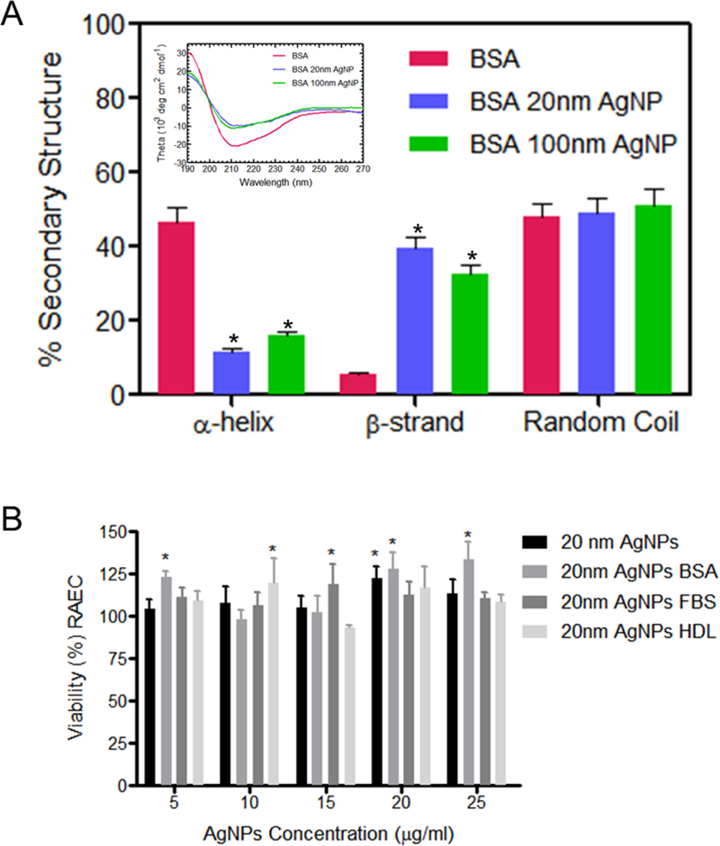
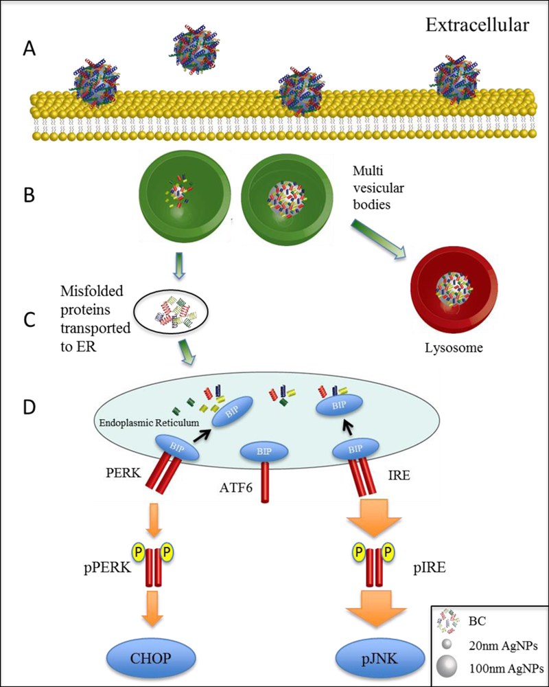
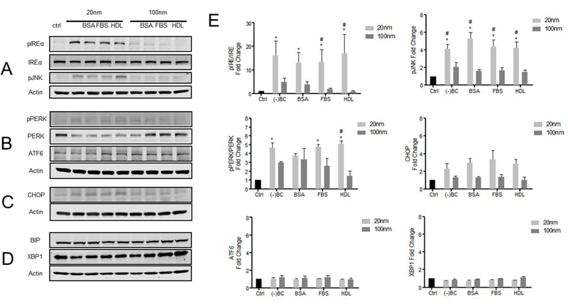
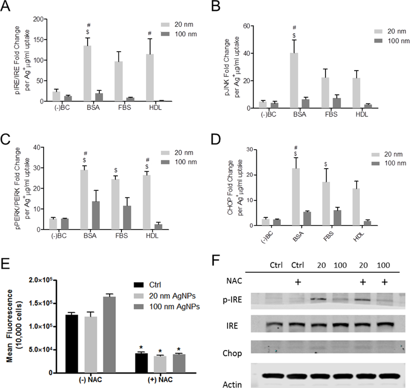
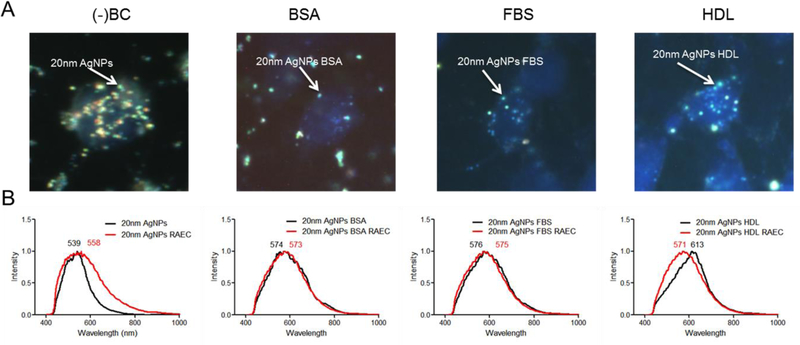
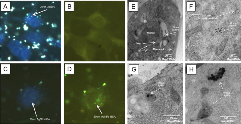
Prior research has demonstrated cells exposed to silver nanoparticles (AgNPs) undergo endoplasmic reticulum (ER) stress leading to cellular apoptosis and toxicity, however, the fundamental mechanism underlying AgNP-induced ER stress is unknown. We hypothesize the biophysical interactions between AgNPs and adsorbed proteins lead to misfolded proteins to elicit an ER stress response. Our investigation examined rat aortic endothelial cells (RAEC) exposed to 20 or 100 nm AgNPs with or without a biocorona (BC) consisting of bovine serum albumin (BSA), high density lipoprotein (HDL) or fetal bovine serum (FBS) to form a complex BC. The presence of a BC consisting of BSA or FBS proteins significantly reduced uptake of 20 nm and 100 nm AgNPs in RAEC. Western blot analysis indicated robust activation of the IREα and PERK pathways in RAEC exposed to 20 nm despite the reduction in uptake by the presence of a BC. This was not observed for the 100 nm AgNPs. Hyperspectral darkfield microscopy qualitatively confirmed that the preformed BC was maintained following uptake by RAEC. Transmission electron microscopy demonstrated a size dependent effect on the sub-cellular localization of AgNPs. Overall, these results suggest that AgNP size, surface area and BC formation governs the induction of ER stress and alterations in intracellular trafficking.

摘要

先前的研究表明,暴露于银纳米粒子(AgNPs)的细胞会经历内质网(ER)应激,导致细胞凋亡和毒性,然而,AgNP 诱导的 ER 应激的基本机制尚不清楚。我们假设 AgNPs 与吸附蛋白之间的生物物理相互作用导致错误折叠的蛋白质引发 ER 应激反应。我们的研究检查了暴露于 20 或 100nm AgNPs 的大鼠主动脉内皮细胞(RAEC),以及是否存在由牛血清白蛋白(BSA)、高密度脂蛋白(HDL)或胎牛血清(FBS)组成的生物被膜(BC),以形成复杂的 BC。BSA 或 FBS 蛋白组成的 BC 的存在显著降低了 RAEC 对 20nm 和 100nm AgNPs 的摄取。Western blot 分析表明,尽管存在 BC 会减少摄取,但暴露于 20nm AgNPs 的 RAEC 中 IREα 和 PERK 途径的活性得到了强烈激活。对于 100nm AgNPs,则没有观察到这种情况。高光谱暗场显微镜定性证实了预形成的 BC 在被 RAEC 摄取后得以维持。透射电子显微镜显示,AgNPs 的大小对其亚细胞定位有尺寸依赖性的影响。总的来说,这些结果表明,AgNP 的尺寸、表面积和 BC 的形成决定了 ER 应激的诱导和细胞内运输的改变。

https://cdn.ncbi.nlm.nih.gov/pmc/blobs/9ae6/6331260/5132fbd718bf/nihms-1515863-f0007.jpg
https://cdn.ncbi.nlm.nih.gov/pmc/blobs/9ae6/6331260/3f25e1c27ac9/nihms-1515863-f0001.jpg
https://cdn.ncbi.nlm.nih.gov/pmc/blobs/9ae6/6331260/aacc2dec26bd/nihms-1515863-f0003.jpg
https://cdn.ncbi.nlm.nih.gov/pmc/blobs/9ae6/6331260/96df7159c911/nihms-1515863-f0004.jpg
https://cdn.ncbi.nlm.nih.gov/pmc/blobs/9ae6/6331260/dd6b314e78b0/nihms-1515863-f0005.jpg
https://cdn.ncbi.nlm.nih.gov/pmc/blobs/9ae6/6331260/81b61af4fd17/nihms-1515863-f0006.jpg
https://cdn.ncbi.nlm.nih.gov/pmc/blobs/9ae6/6331260/5132fbd718bf/nihms-1515863-f0007.jpg

相似文献

1
Biocorona formation contributes to silver nanoparticle induced endoplasmic reticulum stress.生物 corona 形成导致银纳米颗粒诱导的内质网应激。
Ecotoxicol Environ Saf. 2019 Apr 15;170:77-86. doi: 10.1016/j.ecoenv.2018.11.107. Epub 2018 Dec 4.
2
Formation of a protein corona on silver nanoparticles mediates cellular toxicity via scavenger receptors.银纳米颗粒上蛋白质冠的形成通过清道夫受体介导细胞毒性。
Toxicol Sci. 2015 Jan;143(1):136-46. doi: 10.1093/toxsci/kfu217. Epub 2014 Oct 17.
3
A hyperspectral and toxicological analysis of protein corona impact on silver nanoparticle properties, intracellular modifications, and macrophage activation.蛋白质冠层对银纳米颗粒性质、细胞内修饰和巨噬细胞激活影响的高光谱和毒理学分析。
Int J Nanomedicine. 2015 Oct 13;10:6509-21. doi: 10.2147/IJN.S92570. eCollection 2015.
4
Involvement of endoplasmic reticulum stress response and IRE1-mediated ASK1/JNK/Mcl-1 pathways in silver nanoparticle-induced apoptosis of human retinal pigment epithelial cells.内质网应激反应和 IRE1 介导的 ASK1/JNK/Mcl-1 通路参与了银纳米颗粒诱导的人视网膜色素上皮细胞凋亡。
Toxicology. 2020 Sep;442:152540. doi: 10.1016/j.tox.2020.152540. Epub 2020 Jul 24.
5
Properties of silver nanoparticles influencing their uptake in and toxicity to the earthworm Lumbricus rubellus following exposure in soil.影响纳米银颗粒在土壤中被蚯蚓(Lumbricus rubellus)摄取及其毒性的性质。
Environ Pollut. 2016 Nov;218:870-878. doi: 10.1016/j.envpol.2016.08.016. Epub 2016 Aug 11.
6
Endoplasmic reticulum stress signaling is involved in silver nanoparticles-induced apoptosis.内质网应激信号参与了银纳米粒子诱导的细胞凋亡。
Int J Biochem Cell Biol. 2012 Jan;44(1):224-32. doi: 10.1016/j.biocel.2011.10.019. Epub 2011 Nov 3.
7
Silver nanoparticles induce endoplasmatic reticulum stress response in zebrafish.银纳米粒子诱导斑马鱼内质网应激反应。
Toxicol Appl Pharmacol. 2013 Oct 15;272(2):519-28. doi: 10.1016/j.taap.2013.06.011. Epub 2013 Jun 22.
8
Silver nanoparticles induce SH-SY5Y cell apoptosis via endoplasmic reticulum- and mitochondrial pathways that lengthen endoplasmic reticulum-mitochondria contact sites and alter inositol-3-phosphate receptor function.银纳米颗粒通过内质网和线粒体途径诱导SH-SY5Y细胞凋亡,这些途径会延长内质网-线粒体接触位点并改变肌醇-3-磷酸受体功能。
Toxicol Lett. 2018 Mar 15;285:156-167. doi: 10.1016/j.toxlet.2018.01.004. Epub 2018 Jan 3.
9
Response of platelets to silver nanoparticles designed with different surface functionalization.不同表面功能化设计的银纳米颗粒对血小板的响应。
J Inorg Biochem. 2021 Nov;224:111565. doi: 10.1016/j.jinorgbio.2021.111565. Epub 2021 Aug 8.
10
Silver nanoparticles activate endoplasmic reticulum stress signaling pathway in cell and mouse models: The role in toxicity evaluation.银纳米颗粒在细胞和小鼠模型中激活内质网应激信号通路:在毒性评估中的作用。
Biomaterials. 2015 Aug;61:307-15. doi: 10.1016/j.biomaterials.2015.05.029. Epub 2015 May 19.

引用本文的文献

1
Analyzing Molecular Determinants of Nanodrugs' Cytotoxic Effects.分析纳米药物细胞毒性作用的分子决定因素。
Int J Mol Sci. 2025 Jul 11;26(14):6687. doi: 10.3390/ijms26146687.
2
Comprehensive insights into mechanism of nanotoxicity, assessment methods and regulatory challenges of nanomedicines.对纳米毒性机制、纳米药物评估方法及监管挑战的全面洞察。
Discov Nano. 2024 Oct 4;19(1):165. doi: 10.1186/s11671-024-04118-1.
3
Adverse Responses following Exposure to Subtoxic Concentrations of Zinc Oxide and Nickle Oxide Nanoparticles in the Raw 264.7 Cells.

本文引用的文献

1
Gold and silver nanoparticle interactions with human proteins: impact and implications in biocorona formation.金和银纳米颗粒与人类蛋白质的相互作用:生物冠形成中的影响及意义。
J Mater Chem B. 2015 Mar 14;3(10):2075-2082. doi: 10.1039/c4tb01926a. Epub 2015 Feb 2.
2
The Intracellular Destiny of the Protein Corona: A Study on its Cellular Internalization and Evolution.蛋白冠的细胞内命运:对其细胞内化和演变的研究。
ACS Nano. 2016 Nov 22;10(11):10471-10479. doi: 10.1021/acsnano.6b06411. Epub 2016 Nov 2.
3
Protein corona over silver nanoparticles triggers conformational change of proteins and drop in bactericidal potential of nanoparticles: Polyethylene glycol capping as preventive strategy.
在Raw 264.7细胞中暴露于亚毒性浓度的氧化锌和氧化镍纳米颗粒后的不良反应。
Toxics. 2023 Aug 6;11(8):674. doi: 10.3390/toxics11080674.
4
Polystyrene Microplastics Induced Ovarian Toxicity in Juvenile Rats Associated with Oxidative Stress and Activation of the PERK-eIF2α-ATF4-CHOP Signaling Pathway.聚苯乙烯微塑料诱导幼鼠卵巢毒性与氧化应激及PERK-eIF2α-ATF4-CHOP信号通路激活有关。
Toxics. 2023 Feb 27;11(3):225. doi: 10.3390/toxics11030225.
5
Modulation of Pulmonary Toxicity in Metabolic Syndrome Due to Variations in Iron Oxide Nanoparticle-Biocorona Composition.由于氧化铁纳米颗粒-生物冠层组成的变化对代谢综合征中肺毒性的调节作用。
Nanomaterials (Basel). 2022 Jun 11;12(12):2022. doi: 10.3390/nano12122022.
6
Optical Microscopy Systems for the Detection of Unlabeled Nanoparticles.用于检测未标记纳米粒子的光学显微镜系统。
Int J Nanomedicine. 2022 May 13;17:2139-2163. doi: 10.2147/IJN.S355007. eCollection 2022.
7
Functionalized Nanoplastics (NPs) Increase the Toxicity of Metals in Fish Cell Lines.功能化纳米塑料 (NPs) 增加了鱼类细胞系中金属的毒性。
Int J Mol Sci. 2021 Jul 1;22(13):7141. doi: 10.3390/ijms22137141.
8
Insights into Characterization Methods and Biomedical Applications of Nanoparticle-Protein Corona.纳米颗粒-蛋白质冠层的表征方法及生物医学应用洞察
Materials (Basel). 2020 Jul 10;13(14):3093. doi: 10.3390/ma13143093.
9
Continued Efforts on Nanomaterial-Environmental Health and Safety Is Critical to Maintain Sustainable Growth of Nanoindustry.持续努力研究纳米材料的环境健康与安全对于维持纳米产业的可持续增长至关重要。
Small. 2020 May;16(21):e2000603. doi: 10.1002/smll.202000603. Epub 2020 Apr 27.
10
Tackling Complex Analytical Tasks: An ISO/TS-Based Validation Approach for Hydrodynamic Chromatography Single Particle Inductively Coupled Plasma Mass Spectrometry.应对复杂分析任务:基于ISO/TS的流体动力学色谱-单颗粒电感耦合等离子体质谱验证方法
Materials (Basel). 2020 Mar 22;13(6):1447. doi: 10.3390/ma13061447.
银纳米颗粒上的蛋白质冠层引发蛋白质构象变化并降低纳米颗粒的杀菌潜力:聚乙二醇封端作为预防策略。
Colloids Surf B Biointerfaces. 2016 Oct 1;146:577-84. doi: 10.1016/j.colsurfb.2016.06.050. Epub 2016 Jun 28.
4
Colloidal Instability Fosters Agglomeration of Subvisible Particles Created by Rupture of Gels of a Monoclonal Antibody Formed at Silicone Oil-Water Interfaces.胶体不稳定性促进了由硅油 - 水界面处形成的单克隆抗体凝胶破裂产生的亚可见颗粒的聚集。
J Pharm Sci. 2016 Aug;105(8):2338-48. doi: 10.1016/j.xphs.2016.06.010. Epub 2016 Jul 13.
5
A hyperspectral and toxicological analysis of protein corona impact on silver nanoparticle properties, intracellular modifications, and macrophage activation.蛋白质冠层对银纳米颗粒性质、细胞内修饰和巨噬细胞激活影响的高光谱和毒理学分析。
Int J Nanomedicine. 2015 Oct 13;10:6509-21. doi: 10.2147/IJN.S92570. eCollection 2015.
6
Silver nanoparticles activate endoplasmic reticulum stress signaling pathway in cell and mouse models: The role in toxicity evaluation.银纳米颗粒在细胞和小鼠模型中激活内质网应激信号通路:在毒性评估中的作用。
Biomaterials. 2015 Aug;61:307-15. doi: 10.1016/j.biomaterials.2015.05.029. Epub 2015 May 19.
7
Formation of a protein corona on silver nanoparticles mediates cellular toxicity via scavenger receptors.银纳米颗粒上蛋白质冠的形成通过清道夫受体介导细胞毒性。
Toxicol Sci. 2015 Jan;143(1):136-46. doi: 10.1093/toxsci/kfu217. Epub 2014 Oct 17.
8
Immunotoxicity of silver nanoparticles in an intravenous 28-day repeated-dose toxicity study in rats.大鼠静脉内28天重复剂量毒性研究中银纳米颗粒的免疫毒性
Part Fibre Toxicol. 2014 May 7;11:21. doi: 10.1186/1743-8977-11-21.
9
Partial unfolding of a monoclonal antibody: role of a single domain in driving protein aggregation.单克隆抗体的部分去折叠:单个结构域在驱动蛋白质聚集过程中的作用
Biochemistry. 2014 May 27;53(20):3367-77. doi: 10.1021/bi5002163. Epub 2014 May 15.
10
Fast intracellular dissolution and persistent cellular uptake of silver nanoparticles in CHO-K1 cells: implication for cytotoxicity.银纳米颗粒在CHO-K1细胞中的快速细胞内溶解和持续细胞摄取:对细胞毒性的影响。
Nanotoxicology. 2015 Mar;9(2):181-9. doi: 10.3109/17435390.2014.907457. Epub 2014 Apr 16.