Braza Matthew E, Young Jonathon, Hammeke Thomas A, Robison Scott E, Han Dennis P, Warren Clinton C, Carroll Joseph, Stepien Kimberly E
Department of Ophthalmology & Visual Sciences, Medical College of Wisconsin, Milwaukee, Wisconsin, USA.
Department of Cell Biology, Neurobiology and Anatomy, Medical College of Wisconsin, Milwaukee, Wisconsin, USA.
BMJ Open Ophthalmol. 2018 Nov 24;3(1):e000104. doi: 10.1136/bmjophth-2017-000104. eCollection 2018.
Previous work using adaptive optics scanning light ophthalmoscopy (AOSLO) imaging has shown photoreceptor disruption to be a common finding in head and ocular trauma patients. Here an expanded trauma population was examined using a novel imaging technique, split-detector AOSLO, to assess remnant cone structure in areas with significant disruption on confocal AOSLO imaging and to follow photoreceptor changes longitudinally.
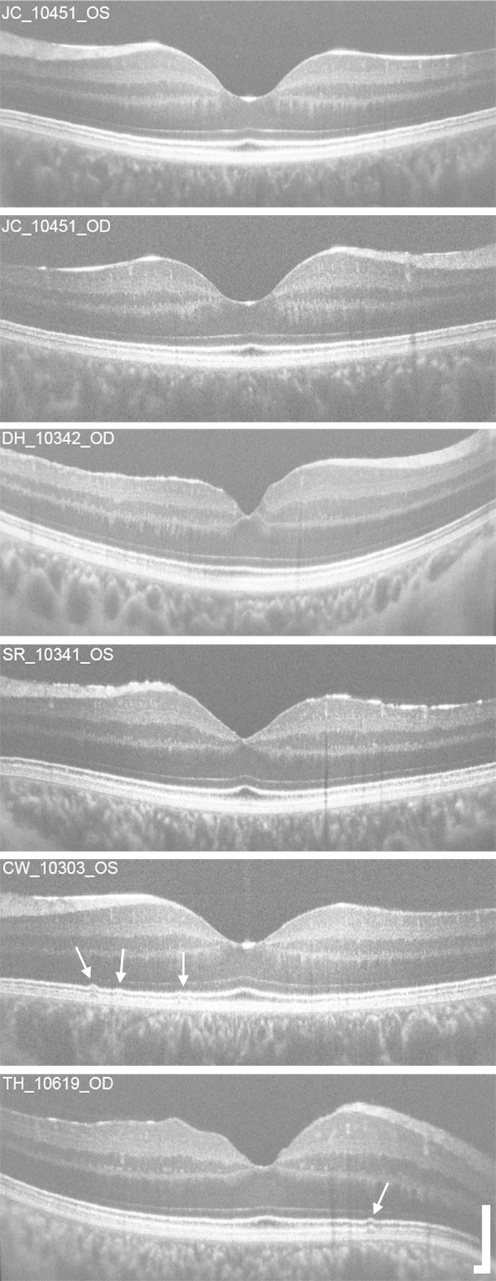
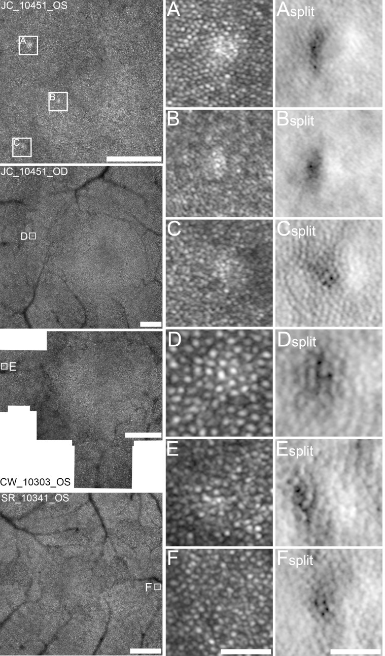
Eight eyes from seven subjects with head and/or ocular trauma underwent imaging with spectral domain optical coherence tomography, confocal AOSLO and split-detector AOSLO to assess foveal and parafoveal photoreceptor structure.
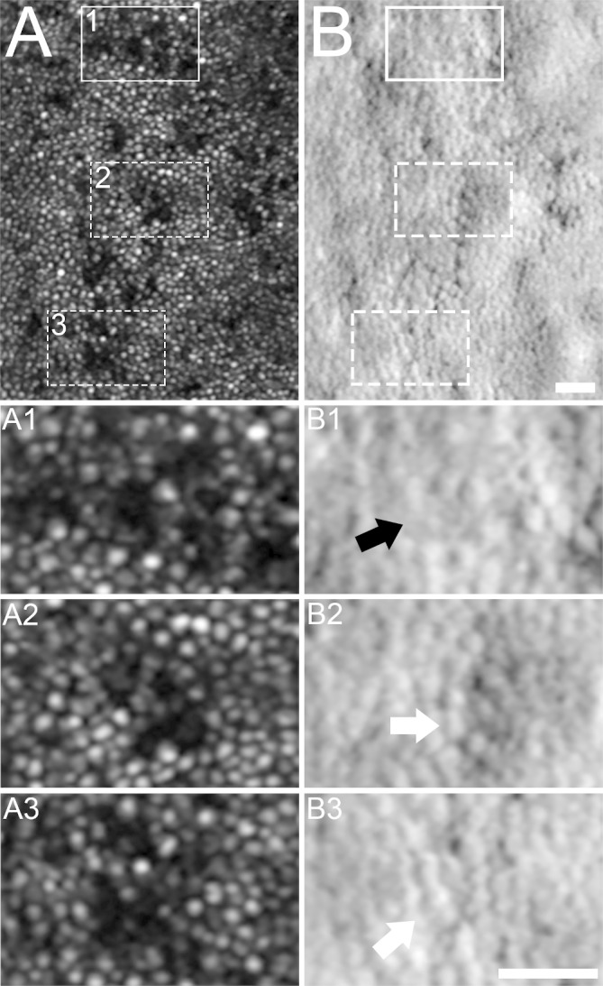
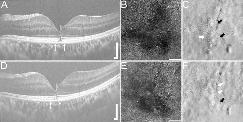
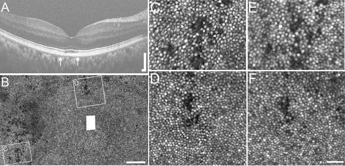
Confocal AOSLO imaging revealed hyporeflective foveal regions in two of eight eyes. Split-detector imaging within the hyporeflective confocal areas showed both remnant and absent inner-segment structure. Both of these eyes were imaged longitudinally and showed variation of the photoreceptor mosaic over time. Four other eyes demonstrated subclinical regions of abnormal waveguiding photoreceptors on multimodal AOSLO imagery but were otherwise normal. Two eyes demonstrated normal foveal cone packing without disruption.
Multimodal imaging can detect subtle photoreceptor abnormalities not necessarily detected by conventional clinical imaging. The addition of split-detector AOSLO revealed the variable condition of inner segments within confocal photoreceptor disruption, confirming the usefulness of dual-modality AOSLO imaging in assessing photoreceptor structure and integrity. Longitudinal imaging demonstrated the dynamic nature of the photoreceptor mosaic after trauma. Multimodal imaging with dual-modality AOSLO improves understanding of visual symptoms and photoreceptor structure changes in patients with head and ocular trauma.
以往使用自适应光学扫描光检眼镜(AOSLO)成像的研究表明,光感受器破坏在头部和眼部创伤患者中是常见现象。在此,我们使用一种新型成像技术——分裂探测器AOSLO,对更多创伤患者进行检查,以评估共焦AOSLO成像显示有明显破坏区域的残余视锥结构,并纵向跟踪光感受器的变化。
对7名患有头部和/或眼部创伤的受试者的8只眼睛进行了光谱域光学相干断层扫描、共焦AOSLO和分裂探测器AOSLO成像,以评估中央凹和中央凹旁的光感受器结构。
共焦AOSLO成像显示8只眼中有2只眼的中央凹区域反射减弱。在反射减弱的共焦区域内进行的分裂探测器成像显示既有残余的内节结构,也有无内节结构的区域。对这两只眼睛都进行了纵向成像,结果显示光感受器镶嵌随着时间而变化。另外4只眼睛在多模态AOSLO图像上显示出波导光感受器异常的亚临床区域,但其他方面正常。2只眼睛显示中央凹视锥细胞排列正常,未受破坏。
多模态成像能够检测出传统临床成像不一定能检测到的细微光感受器异常。添加分裂探测器AOSLO揭示了共焦光感受器破坏区域内可变的内节状况,证实了双模态AOSLO成像在评估光感受器结构和完整性方面的有用性。纵向成像显示了创伤后光感受器镶嵌的动态性质。双模态AOSLO的多模态成像有助于更好地理解头部和眼部创伤患者的视觉症状和光感受器结构变化。