Ly Angelica, Phu Jack, Katalinic Paula, Kalloniatis Michael
Centre for Eye Health, The University of New South Wales, Sydney, New South Wales, Australia.
Faculty of Science, School of Optometry and Vision Science, The University of New South Wales, Sydney, New South Wales, Australia.
Clin Exp Optom. 2019 May;102(3):242-259. doi: 10.1111/cxo.12847. Epub 2018 Dec 17.
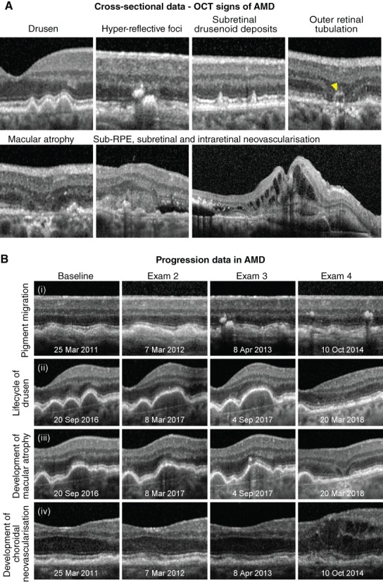
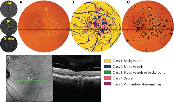
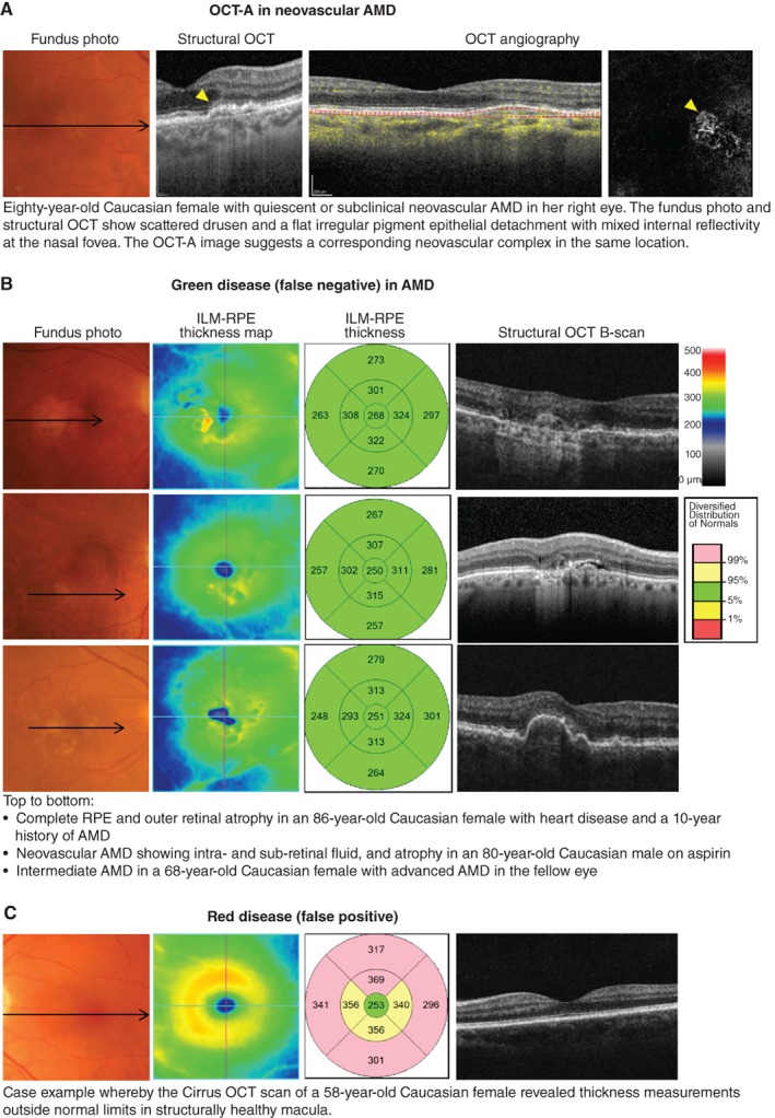
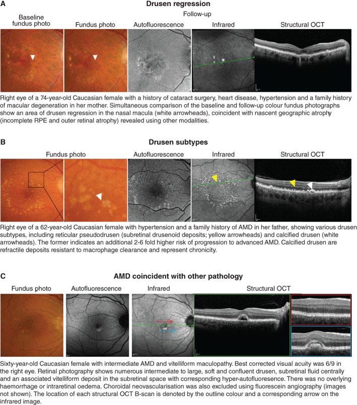
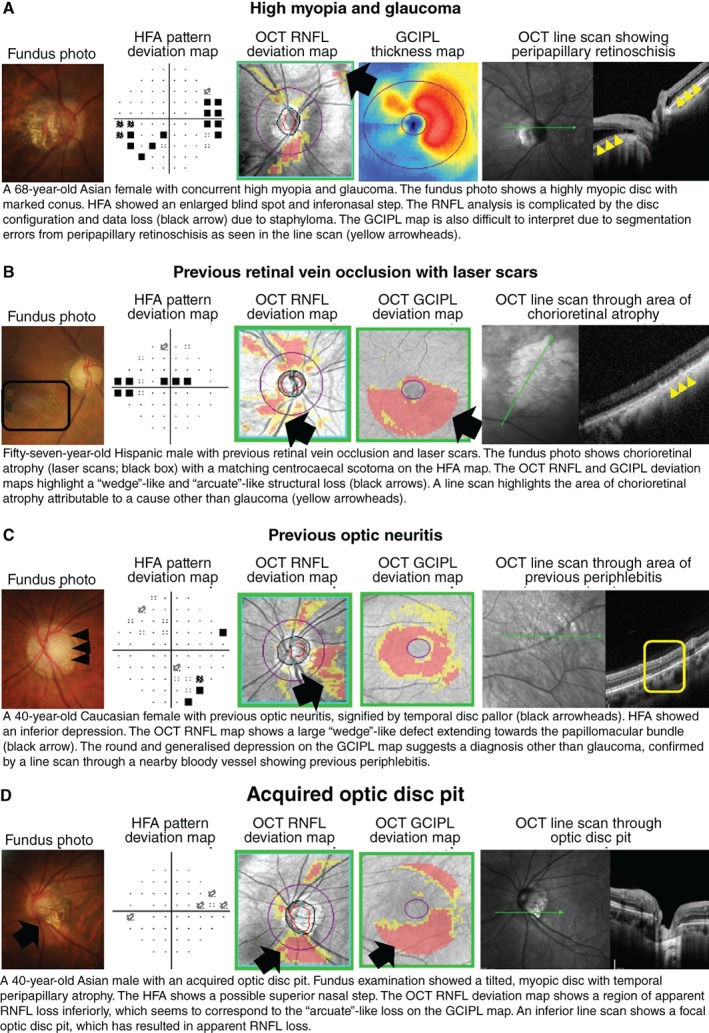
Optical coherence tomography is an imaging technology that has revolutionised the detection, assessment and management of ocular disease. It is now a mainstream technology in clinical practice and is performed by non-specialised personnel in some settings. This article provides a clinical perspective on the implications of that movement and describes best practice using multimodal imaging and an evidence-based approach. Practical, illustrative guides on the interpretation of optical coherence tomography are provided for three major diseases of the ocular fundus, in which optical coherence tomography is often crucial to management: age-related macular degeneration, diabetic retinopathy and glaucoma. Topics discussed include: cross-sectional and longitudinal signs in ocular disease, so-called 'red-green' disease whereby clinicians rely on machine/statistical comparisons for diagnosis in managing treatment-naïve patients, and the utility of optical coherence tomography angiography and machine learning.
光学相干断层扫描是一项彻底改变眼部疾病检测、评估和管理方式的成像技术。它现已成为临床实践中的主流技术,在某些情况下由非专业人员操作。本文从临床角度阐述了这一技术发展的影响,并介绍了使用多模态成像和循证方法的最佳实践。针对眼部三大主要疾病(在这些疾病中,光学相干断层扫描对治疗管理往往至关重要:年龄相关性黄斑变性、糖尿病视网膜病变和青光眼),提供了光学相干断层扫描解读的实用说明指南。讨论的主题包括:眼部疾病的横断面和纵向体征、所谓的“红绿色”疾病(即临床医生在管理初治患者时依靠机器/统计比较进行诊断),以及光学相干断层扫描血管造影和机器学习的效用。