Phu Jack, Wang Henrietta, Miao Sephora, Zhou Lydia, Khuu Sieu K, Kalloniatis Michael
Centre for Eye Health, University of New South Wales, Kensington, New South Wales, Australia.
School of Optometry and Vision Science, University of New South Wales, Kensington, New South Wales, Australia *
Optom Vis Sci. 2018 Oct;95(10):959-970. doi: 10.1097/OPX.0000000000001286.
We demonstrate that the visual field defects in patients with tilted disc syndrome can be reduced or eliminated by neutralizing the peripheral scotoma in the area of posterior retinal bowing, which may allow differentiation between a congenital anomaly and acquired pathology.
Tilted disc syndrome is a congenital and unchanging condition that may present with visual field defects mimicking loss seen in neurological diseases, such as transsynaptic retrograde degeneration. Our purpose was to systematically investigate the ability of a neutralized peripheral refraction to eliminate refractive visual field defects seen in tilted disc syndrome. This was compared with the same technique performed on patients with neurological deficits.
The Humphrey Field Analyzer was used to measure sensitivities across the 30-2 test grid in 14 patients with tilted disc syndrome using four refractive corrections: habitual near correction and with an additional -1.00, -2.00 or -3.00 D negative lens added as correction lenses. Peripheral refractive errors along the horizontal meridian were determined using peripheral retinoscopy and thus allowed calculation of residual peripheral refraction with different levels of refractive correction. Visual field defects were assessed qualitatively and quantitatively using sensitivities and probability scores in both patient groups.
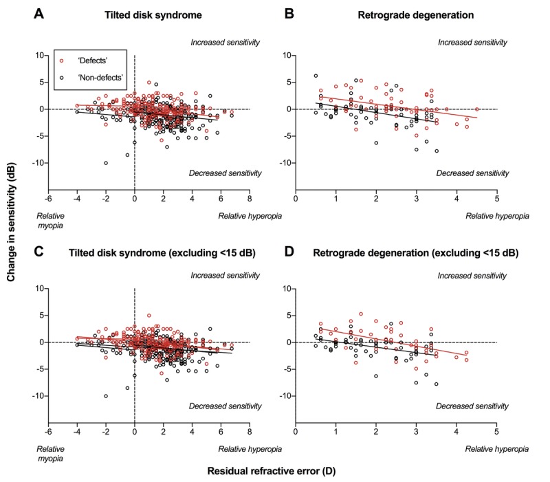
A smaller residual refractive error after the application of negative addition lenses correlated with improvement in visual field defects in terms of sensitivity and probability scores in patients with tilted disc syndrome. Patients with established neurological deficits (retrograde degeneration) showed improvement in sensitivities but not in probability scores.
Neutralizing the refractive error at the region of posterior retinal bowing due to tilted disc syndrome reduces the apparent visual field defect. This may be a useful and rapid test to help differentiate between tilted disc syndrome and other pathological causes of visual field defects such as neurological deficits.
我们证明,通过中和后极视网膜弓区域的周边暗点,可减少或消除倾斜盘综合征患者的视野缺损,这可能有助于区分先天性异常和后天性病变。
倾斜盘综合征是一种先天性且不变的病症,可能出现类似神经疾病(如跨突触逆行性变性)中所见视野缺损的症状。我们的目的是系统研究中和周边屈光不正以消除倾斜盘综合征中所见屈光性视野缺损的能力。并将其与对神经功能缺损患者进行的相同技术进行比较。
使用汉弗莱视野分析仪,对14例倾斜盘综合征患者在30-2测试网格上进行敏感度测量,采用四种屈光矫正方式:习惯性近矫正以及额外添加-1.00、-2.00或-3.00D负透镜作为矫正镜片。使用周边视网膜镜检查确定沿水平子午线的周边屈光不正,从而计算不同屈光矫正水平下的残余周边屈光不正。使用敏感度和概率分数对两组患者的视野缺损进行定性和定量评估。
对于倾斜盘综合征患者,应用负球镜附加镜片后残余屈光不正较小与视野缺损在敏感度和概率分数方面的改善相关。已确诊神经功能缺损(逆行性变性)的患者敏感度有所改善,但概率分数未改善。
中和因倾斜盘综合征导致的后极视网膜弓区域的屈光不正可减少明显的视野缺损。这可能是一种有用且快速的检查方法,有助于区分倾斜盘综合征和其他导致视野缺损的病理原因,如神经功能缺损。