Department of Neurology, Alzheimer Center Amsterdam, Amsterdam Neuroscience, Vrije Universiteit Amsterdam, Amsterdam UMC, Mailbox 7057, Amsterdam, 1007 MB, The Netherlands.
Department of Pathology, Amsterdam Neuroscience, Amsterdam UMC, Vrije Universiteit Amsterdam, Amsterdam, The Netherlands.
Acta Neuropathol Commun. 2018 Dec 28;6(1):147. doi: 10.1186/s40478-018-0650-x.
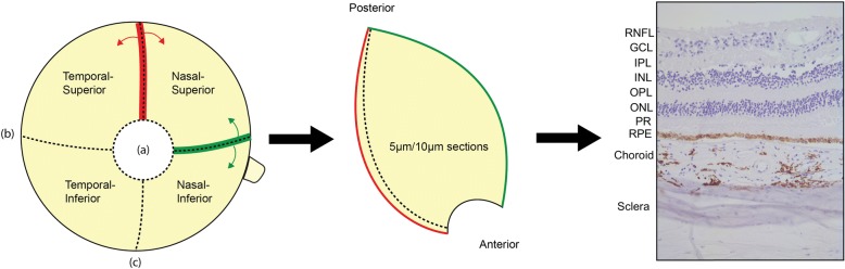
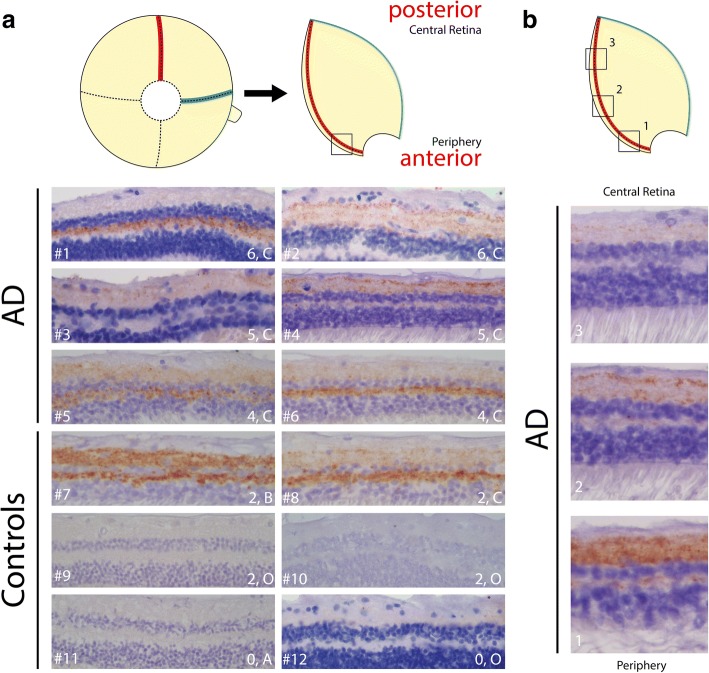
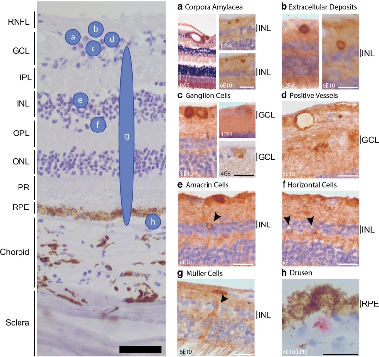
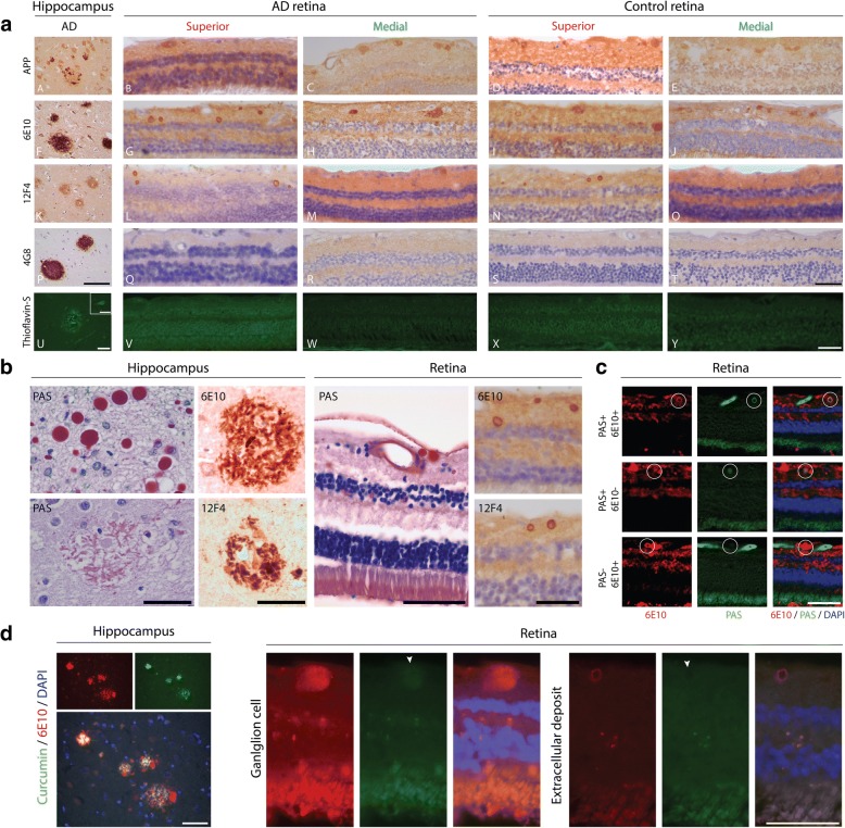
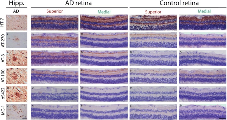
In-vivo labeling of retinal amyloid-beta(Aβ) and tau has potential as non-invasive biomarker for Alzheimer's disease (AD). However, literature on the presence of Aβ and phosphorylated tau (pTau) in AD retinas is inconclusive. We therefore assessed the presence of Aβ and pTau in post-mortem retinas in 6 AD and 6 control cases who donated brains and eyes to the Netherlands Brain Bank. Neuropathological diagnosis of AD was made according to NIA-AA criteria. Formalin fixed retinas were dissected in quadrants and cross-sections of medial and superior retinas were made. Immuno-histochemical stainings were performed for Aβ, amyloid precursor protein (APP) and pTau. To assess translation to an in-vivo set up using curcumin as labelling fluorophore, co-stainings with curcumin were performed. No typical Aβ-plaques and neurofibrillary tangles, like in the cerebral cortex, were observed in AD retinas. A diffuse immunoreactive signal for pTau was increased in the inner and outer plexiform layers of the retina in AD cases compared to control cases with absence of cerebral amyloid pathology. Immunostaining with anti-Aβ and anti-APP antibodies yielded signal in ganglion cells, amacrine cells, horizontal cells and Müller cells in both control and AD cases. We observed small extracellular deposits positive for anti-Aβ antibodies 12F4 and 6E10 and negative for 4G8 and curcumin. A subset of these deposits could be characterized as corpora amylacea. In conclusion we found that retinal manifestations of AD pathology appear to be different compared to cerebral AD pathology. Using a qualitative cross-sectional approach, we did not find Aβ/APP related differences in the retina between AD and control subjects. In contrast, tau related changes were found to be present in cases with cerebral AD pathology, suggesting retinal tau as a potential biomarker for AD.
在体标记视网膜淀粉样β(Aβ)和tau 有望成为阿尔茨海默病(AD)的非侵入性生物标志物。然而,关于 AD 视网膜中 Aβ 和磷酸化 tau(pTau)的存在的文献尚无定论。因此,我们评估了 6 例 AD 和 6 例对照病例死后视网膜中 Aβ 和 pTau 的存在情况,这些病例向荷兰脑库捐献了大脑和眼睛。AD 的神经病理学诊断根据 NIA-AA 标准进行。福尔马林固定的视网膜被切成四部分,制作了内侧和上部视网膜的横截面。进行了 Aβ、淀粉样前体蛋白(APP)和 pTau 的免疫组织化学染色。为了评估使用姜黄素作为标记荧光团的体内设置的转化,进行了与姜黄素的共染色。在 AD 视网膜中未观察到典型的 Aβ 斑块和神经原纤维缠结,如大脑皮质中的那样。与无大脑淀粉样蛋白病理的对照病例相比,AD 病例的视网膜内、外丛状层中的 pTau 免疫反应性信号增加。抗 Aβ 和抗 APP 抗体的免疫染色在对照和 AD 病例中均在神经节细胞、无长突细胞、水平细胞和 Müller 细胞中产生信号。我们观察到小的细胞外沉积物对 12F4 和 6E10 抗 Aβ 抗体呈阳性,对 4G8 和姜黄素呈阴性。这些沉积物中的一部分可以被描述为淀粉样体。总之,我们发现 AD 病理学的视网膜表现似乎与大脑 AD 病理学不同。使用定性的横截面方法,我们在 AD 和对照受试者的视网膜中没有发现 Aβ/APP 相关的差异。相比之下,在有大脑 AD 病理的病例中发现了 tau 相关的变化,提示视网膜 tau 可能是 AD 的潜在生物标志物。