Curr Opin Rheumatol. 2019 Mar;31(2):134-143. doi: 10.1097/BOR.0000000000000582.
The present review addresses diagnostic methods for crystalline arthritis including synovial fluid analysis, ultrasound, and dual energy CT scan (DECT).
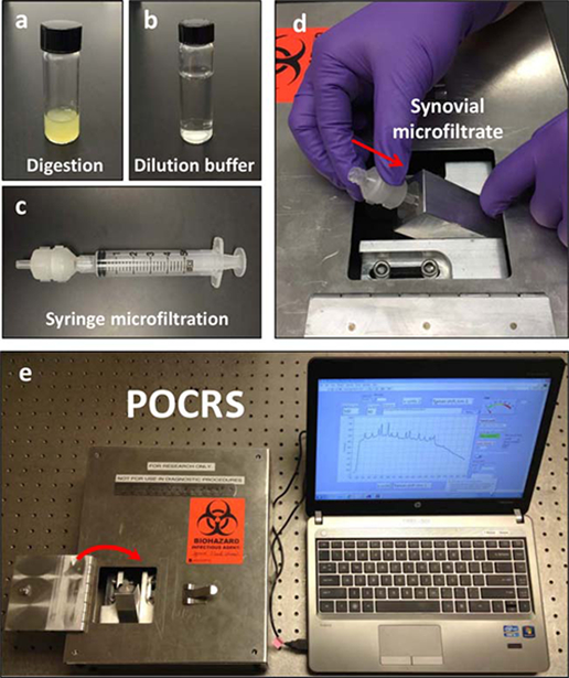
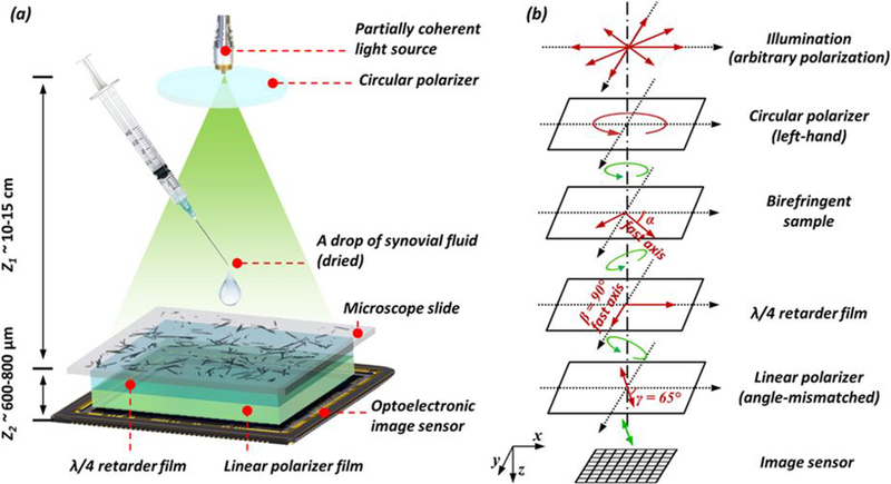
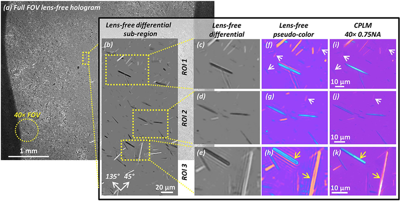
There are new technologies on the horizon to improve the ease, sensitivity, and specificity of synovial fluid analysis. Raman spectroscopy uses the spectral signature that results from a material's unique energy absorption and scatter for crystal identification. Lens-free microscopy directly images synovial fluid aspirate on to a complementary metal-oxide semiconductor chip, providing a high-resolution, wide field of view (∼20 mm) image. Raman spectroscopy and lens-free microscopy may provide additional benefit over compensated polarized light microscopy synovial fluid analysis by quantifying crystal density in synovial fluid samples. Ultrasound and DECT have good sensitivity and specificity for the identification of monosodium urate (MSU) and calcium pyrophosphate (CPP) crystals. However, both have limitations in patients with recent onset gout and low urate burdens.
New technologies promise improved methods for detection of MSU and CPP crystals. At this time, limitations of these technologies do not replace the need for synovial fluid aspiration for confirmation of crystal detection. None of these technologies address the often concomitant indication to rule out infectious arthritis.
本综述讨论了晶体性关节炎的诊断方法,包括滑液分析、超声和双能 CT 扫描(DECT)。
为了提高滑液分析的简便性、敏感性和特异性,新技术正在不断涌现。拉曼光谱利用物质独特的能量吸收和散射产生的光谱特征来进行晶体鉴定。无透镜显微镜直接对互补金属氧化物半导体芯片上的滑膜液抽吸物进行成像,提供高分辨率、宽视野(约 20mm)的图像。拉曼光谱和无透镜显微镜可能通过量化滑膜液样本中的晶体密度,为补偿偏光显微镜滑膜液分析提供额外的益处。超声和 DECT 对单钠尿酸盐(MSU)和焦磷酸钙(CPP)晶体的识别具有良好的敏感性和特异性。然而,在尿酸负荷低且痛风发作时间较短的患者中,这两种方法都存在局限性。
新技术有望改善 MSU 和 CPP 晶体的检测方法。目前,这些技术的局限性并没有取代对滑膜液抽吸以确认晶体检测的需求。这些技术都不能解决经常伴随的排除感染性关节炎的指征。