• 文献检索
  • 文档翻译
  • 深度研究
  • 学术资讯
  • Suppr Zotero 插件Zotero 插件
  • 邀请有礼
  • 套餐&价格
  • 历史记录
应用&插件
Suppr Zotero 插件Zotero 插件浏览器插件Mac 客户端Windows 客户端微信小程序
定价
高级版会员购买积分包购买API积分包
服务
文献检索文档翻译深度研究API 文档MCP 服务
关于我们
关于 Suppr公司介绍联系我们用户协议隐私条款
关注我们

Suppr 超能文献

核心技术专利:CN118964589B侵权必究
粤ICP备2023148730 号-1Suppr @ 2026

文献检索

告别复杂PubMed语法,用中文像聊天一样搜索,搜遍4000万医学文献。AI智能推荐,让科研检索更轻松。

立即免费搜索

文件翻译

保留排版,准确专业,支持PDF/Word/PPT等文件格式,支持 12+语言互译。

免费翻译文档

深度研究

AI帮你快速写综述,25分钟生成高质量综述,智能提取关键信息,辅助科研写作。

立即免费体验

数字病理学二十年:走过的道路、未来展望及供应商中立档案的兴起概述

Twenty Years of Digital Pathology: An Overview of the Road Travelled, What is on the Horizon, and the Emergence of Vendor-Neutral Archives.

作者信息

Pantanowitz Liron, Sharma Ashish, Carter Alexis B, Kurc Tahsin, Sussman Alan, Saltz Joel

机构信息

Department of Pathology, University of Pittsburgh Medical Center, Pittsburgh, PA, USA.

Department of Biomedical Informatics, Emory University, GA, USA.

出版信息

J Pathol Inform. 2018 Nov 21;9:40. doi: 10.4103/jpi.jpi_69_18. eCollection 2018.

DOI:10.4103/jpi.jpi_69_18
PMID:30607307
原文链接:https://pmc.ncbi.nlm.nih.gov/articles/PMC6289005/
Abstract

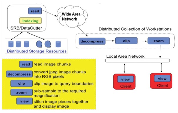
Almost 20 years have passed since the commercial introduction of whole-slide imaging (WSI) scanners. During this time, the creation of various WSI devices with the ability to digitize an entire glass slide has transformed the field of pathology. Parallel advances in computational technology and storage have permitted rapid processing of large-scale WSI datasets. This article provides an overview of important past and present efforts related to WSI. An account of how the virtual microscope evolved from the need to visualize and manage satellite data for earth science applications is provided. The article also discusses important milestones beginning from the first WSI scanner designed by Bacus to the Food and Drug Administration approval of the first digital pathology system for primary diagnosis in surgical pathology. As pathology laboratories commit to going fully digitalize, the need has emerged to include WSIs into an enterprise-level vendor-neutral archive (VNA). The different types of VNAs available are reviewed as well as how best to implement them and how pathology can benefit from participating in this effort. Differences between traditional image algorithms that extract pixel-, object-, and semantic-level features versus deep learning methods are highlighted. The need for large-scale data management, analysis, and visualization in computational pathology is also addressed.

摘要

自全切片成像(WSI)扫描仪商业化推出以来,已过去近20年。在此期间,各种能够将整个玻璃切片数字化的WSI设备的出现改变了病理学领域。计算技术和存储方面的同步进步使得大规模WSI数据集能够得到快速处理。本文概述了与WSI相关的过去和现在的重要工作。介绍了虚拟显微镜如何从地球科学应用中可视化和管理卫星数据的需求演变而来。文章还讨论了从Bacus设计的第一台WSI扫描仪到食品药品监督管理局批准首个用于外科病理学初步诊断的数字病理系统等重要里程碑。随着病理实验室致力于全面数字化,将WSI纳入企业级供应商中立存档(VNA)的需求应运而生。本文回顾了可用的不同类型的VNA,以及如何最好地实施它们,以及病理学如何从参与这项工作中受益。强调了提取像素级、对象级和语义级特征的传统图像算法与深度学习方法之间的差异。还讨论了计算病理学中大规模数据管理、分析和可视化的需求。

https://cdn.ncbi.nlm.nih.gov/pmc/blobs/7746/6289005/7526e2588106/JPI-9-40-g011.jpg
https://cdn.ncbi.nlm.nih.gov/pmc/blobs/7746/6289005/ee557145d5a3/JPI-9-40-g001.jpg
https://cdn.ncbi.nlm.nih.gov/pmc/blobs/7746/6289005/6b01b3a920bd/JPI-9-40-g002.jpg
https://cdn.ncbi.nlm.nih.gov/pmc/blobs/7746/6289005/7edf324b898b/JPI-9-40-g003.jpg
https://cdn.ncbi.nlm.nih.gov/pmc/blobs/7746/6289005/2e9aa1834ff0/JPI-9-40-g004.jpg
https://cdn.ncbi.nlm.nih.gov/pmc/blobs/7746/6289005/6401b60b8133/JPI-9-40-g005.jpg
https://cdn.ncbi.nlm.nih.gov/pmc/blobs/7746/6289005/fc756fa4f7ba/JPI-9-40-g008.jpg
https://cdn.ncbi.nlm.nih.gov/pmc/blobs/7746/6289005/76b2ceeeec54/JPI-9-40-g010.jpg
https://cdn.ncbi.nlm.nih.gov/pmc/blobs/7746/6289005/7526e2588106/JPI-9-40-g011.jpg

相似文献

1
Twenty Years of Digital Pathology: An Overview of the Road Travelled, What is on the Horizon, and the Emergence of Vendor-Neutral Archives.数字病理学二十年:走过的道路、未来展望及供应商中立档案的兴起概述
J Pathol Inform. 2018 Nov 21;9:40. doi: 10.4103/jpi.jpi_69_18. eCollection 2018.
2
Utility of whole slide imaging and virtual microscopy in prostate pathology.全玻片成像和虚拟显微镜在前列腺病理学中的应用。
APMIS. 2012 Apr;120(4):298-304. doi: 10.1111/j.1600-0463.2011.02872.x.
3
Whole Slide Imaging (WSI) in Pathology: Current Perspectives and Future Directions.病理全切片成像(WSI):现状与未来方向。
J Digit Imaging. 2020 Aug;33(4):1034-1040. doi: 10.1007/s10278-020-00351-z.
4
The Importance of eSlide Macro Images for Primary Diagnosis with Whole Slide Imaging.eSlide宏图像在全切片成像初步诊断中的重要性。
J Pathol Inform. 2018 Dec 24;9:46. doi: 10.4103/jpi.jpi_70_18. eCollection 2018.
5
Multilayer outperforms single-layer slide scanning in AI-based classification of whole slide images with low-burden acid-fast mycobacteria (AFB).多层扫描优于单层扫描,可基于人工智能对低负担抗酸分枝杆菌(AFB)全玻片图像进行分类。
Comput Methods Programs Biomed. 2023 Jun;234:107518. doi: 10.1016/j.cmpb.2023.107518. Epub 2023 Mar 28.
6
Applications of discriminative and deep learning feature extraction methods for whole slide image analysis: A survey.判别式和深度学习特征提取方法在全切片图像分析中的应用:一项综述。
J Pathol Inform. 2023 Sep 14;14:100335. doi: 10.1016/j.jpi.2023.100335. eCollection 2023.
7
Whole slide imaging equivalency and efficiency study: experience at a large academic center.全 slides 成像等效性和效率研究:大型学术中心的经验。
Mod Pathol. 2019 Jul;32(7):916-928. doi: 10.1038/s41379-019-0205-0. Epub 2019 Feb 18.
8
Optimized JPEG 2000 Compression for Efficient Storage of Histopathological Whole-Slide Images.用于高效存储组织病理学全切片图像的优化JPEG 2000压缩
J Pathol Inform. 2018 May 25;9:20. doi: 10.4103/jpi.jpi_69_17. eCollection 2018.
9
(Re) Defining the High-Power Field for Digital Pathology.重新定义数字病理学的高倍视野
J Pathol Inform. 2020 Oct 9;11:33. doi: 10.4103/jpi.jpi_48_20. eCollection 2020.
10
FlexTileSource: An OpenSeadragon Extension for Efficient Whole-Slide Image Visualization.FlexTileSource:一种用于高效全切片图像可视化的OpenSeadragon扩展
J Pathol Inform. 2021 Sep 14;12:31. doi: 10.4103/jpi.jpi_13_21. eCollection 2021.

引用本文的文献

1
Innovative approach for the qualitative-quantitative assessment of neurodevelopment biomarkers research in placenta tissue using immunohistochemistry digital image analysis.使用免疫组织化学数字图像分析对胎盘组织中神经发育生物标志物研究进行定性-定量评估的创新方法。
Biol Methods Protoc. 2025 Jul 11;10(1):bpaf056. doi: 10.1093/biomethods/bpaf056. eCollection 2025.
2
An open-source platform for structured annotation and computational workflows in digital pathology research.一个用于数字病理学研究中结构化注释和计算工作流程的开源平台。
Sci Rep. 2025 Aug 7;15(1):28910. doi: 10.1038/s41598-025-13546-7.
3
Whole Slide Imaging (WSI) in Pathology: Emerging Trends and Future Applications in Clinical Diagnostics, Medical Education, and Pathology.

本文引用的文献

1
SparkGIS: Efficient Comparison and Evaluation of Algorithm Results in Tissue Image Analysis Studies.SparkGIS:组织图像分析研究中算法结果的高效比较与评估
Biomed Data Manag Graph Online Querying (2015). 2016;9579:134-146. doi: 10.1007/978-3-319-41576-5_10. Epub 2016 Jun 24.
2
Conventional Microscopical versus Digital Whole-Slide Imaging-Based Diagnosis of Thin-Layer Cervical Specimens: A Validation Study.传统显微镜检查与基于数字全玻片成像的薄层宫颈标本诊断:一项验证研究。
J Pathol Inform. 2018 Aug 27;9:29. doi: 10.4103/jpi.jpi_28_18. eCollection 2018.
3
Center-Focusing Multi-task CNN with Injected Features for Classification of Glioma Nuclear Images.
病理学中的全切片成像(WSI):临床诊断、医学教育和病理学领域的新兴趋势及未来应用
Iran J Pathol. 2025 Summer;20(3):257-265. doi: 10.30699/ijp.2025.2044210.3367. Epub 2025 Jul 1.
4
Development of a national pathology training system using digital pathology and SNOMED-CT.利用数字病理学和SNOMED-CT开发国家病理学培训系统。
J Pathol Inform. 2025 Jun 18;18:100456. doi: 10.1016/j.jpi.2025.100456. eCollection 2025 Aug.
5
Variation within and between digital pathology and light microscopy for the diagnosis of histopathology slides: blinded crossover comparison study.数字病理学与光学显微镜检查在组织病理学切片诊断中的内部及相互间差异:双盲交叉对比研究
Health Technol Assess. 2025 Jul;29(30):1-75. doi: 10.3310/SPLK4325.
6
Development and retrospective validation of an artificial intelligence system for diagnostic assessment of prostate biopsies: study protocol.用于前列腺活检诊断评估的人工智能系统的开发与回顾性验证:研究方案
BMJ Open. 2025 Jul 7;15(7):e097591. doi: 10.1136/bmjopen-2024-097591.
7
[Training in the age of digitalization- evaluation of the digital Münster dermatopathology course].[数字化时代的培训——明斯特数字皮肤病理学课程评估]
Dermatologie (Heidelb). 2025 May 23. doi: 10.1007/s00105-025-05516-x.
8
Perspectives on Reducing Barriers to the Adoption of Digital and Computational Pathology Technology by Clinical Labs.临床实验室减少采用数字和计算病理学技术障碍的相关观点。
Diagnostics (Basel). 2025 Mar 21;15(7):794. doi: 10.3390/diagnostics15070794.
9
Unlocking the potential of digital pathology: Novel baselines for compression.释放数字病理学的潜力:压缩的新基线
J Pathol Inform. 2025 Jan 28;17:100421. doi: 10.1016/j.jpi.2025.100421. eCollection 2025 Apr.
10
An update on applications of digital pathology: primary diagnosis; telepathology, education and research.数字病理学应用的最新进展:初步诊断;远程病理学、教育与研究。
Diagn Pathol. 2025 Feb 12;20(1):17. doi: 10.1186/s13000-025-01610-9.
具有注入特征的中心聚焦多任务卷积神经网络用于胶质瘤细胞核图像分类
IEEE Winter Conf Appl Comput Vis. 2017 Mar;2017:834-841. doi: 10.1109/WACV.2017.98. Epub 2017 May 15.
4
US Food and Drug Administration Approval of Whole Slide Imaging for Primary Diagnosis: A Key Milestone Is Reached and New Questions Are Raised.美国食品和药物管理局批准全玻片成像用于初步诊断:一个关键的里程碑已经达成,同时也引发了新的问题。
Arch Pathol Lab Med. 2018 Nov;142(11):1383-1387. doi: 10.5858/arpa.2017-0496-CP. Epub 2018 Apr 30.
5
Routine Digital Pathology Workflow: The Catania Experience.常规数字病理学工作流程:卡塔尼亚经验
J Pathol Inform. 2017 Dec 19;8:51. doi: 10.4103/jpi.jpi_58_17. eCollection 2017.
6
PanCancer insights from The Cancer Genome Atlas: the pathologist's perspective.从癌症基因组图谱中获得的泛癌症洞察:病理学家视角。
J Pathol. 2018 Apr;244(5):512-524. doi: 10.1002/path.5028. Epub 2018 Feb 22.
7
QuPath: Open source software for digital pathology image analysis.QuPath:用于数字病理学图像分析的开源软件。
Sci Rep. 2017 Dec 4;7(1):16878. doi: 10.1038/s41598-017-17204-5.
8
An Image Analysis Resource for Cancer Research: PIIP-Pathology Image Informatics Platform for Visualization, Analysis, and Management.癌症研究的图像分析资源:用于可视化、分析和管理的PIIP-病理学图像信息学平台。
Cancer Res. 2017 Nov 1;77(21):e83-e86. doi: 10.1158/0008-5472.CAN-17-0323.
9
A Containerized Software System for Generation, Management, and Exploration of Features from Whole Slide Tissue Images.一种用于从全切片组织图像生成、管理和探索特征的容器化软件系统。
Cancer Res. 2017 Nov 1;77(21):e79-e82. doi: 10.1158/0008-5472.CAN-17-0316.
10
The Digital Slide Archive: A Software Platform for Management, Integration, and Analysis of Histology for Cancer Research.数字切片存档:用于癌症研究组织学管理、整合与分析的软件平台。
Cancer Res. 2017 Nov 1;77(21):e75-e78. doi: 10.1158/0008-5472.CAN-17-0629.