Luo Xiaomei, Chen Guanmao, Jia Yanbin, Gong JiaYing, Qiu Shaojuan, Zhong Shuming, Zhao Lianping, Chen Feng, Lai Shunkai, Qi Zhangzhang, Huang Li, Wang Ying
Medical Imaging Center, First Affiliated Hospital of Jinan University, Guangzhou, China.
Institute of Molecular and Functional Imaging, Jinan University, Guangzhou, China.
Front Psychiatry. 2018 Dec 18;9:705. doi: 10.3389/fpsyt.2018.00705. eCollection 2018.
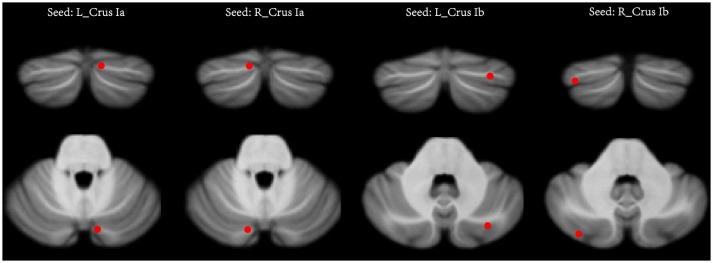
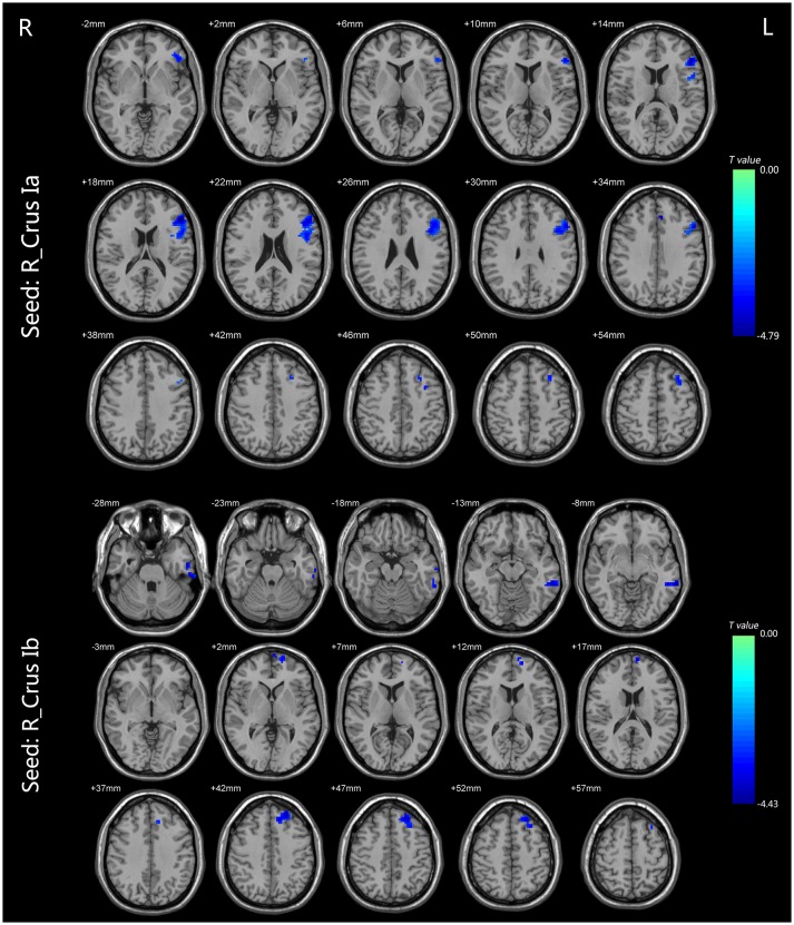
Bipolar disorder (BD) is a common psychiatric disease. Although structural and functional abnormalities of the cerebellum in BD patients have been reported by recent neuroimaging studies, the cerebellar-cerebral functional connectivity (FC) has not yet been examined. The present study aims to investigate the FC between the cerebellum and cerebrum, particularly the central executive network (CEN) and the default-mode network (DMN) in bipolar II disorder (BD II). Ninety-four patients with unmedicated BD II depression and 100 healthy controls (HCs) underwent the resting-state functional magnetic resonance imaging. Seed-based connectivity analyses were performed using cerebellar seeds previously identified as being involved in the CEN (bilateral Crus Ia) and DMN (bilateral Crus Ib). Compared with HCs, BD II depression patients appeared decreased FC in the right Crus Ia-left dorsal lateral prefrontal cortex (dlPFC) and -left anterior cingulate cortex (ACC), the right Crus Ib-left medial prefrontal cortex (mPFC), -left middle temporal gyrus (MTG), and -left inferior temporal gyrus (ITG). No altered FC between the left Crus Ia or Crus Ib and the cerebral regions was found. Patients with BD II depression showed disrupted FC between the cerebellum and the CEN (mainly in the left dlPFC and ACC) and DMN (mainly in the left mPFC and temporal lobe), suggesting the significant role of the cerebellum-CEN and -DMN connectivity in the pathogenesis of BD.
双相情感障碍(BD)是一种常见的精神疾病。尽管最近的神经影像学研究报道了BD患者小脑的结构和功能异常,但尚未对小脑-大脑功能连接(FC)进行研究。本研究旨在调查双相II型障碍(BD II)患者小脑与大脑之间的FC,特别是中央执行网络(CEN)和默认模式网络(DMN)。94例未用药的BD II型抑郁症患者和100名健康对照者(HCs)接受了静息态功能磁共振成像检查。使用先前确定参与CEN(双侧 Crus Ia)和DMN(双侧 Crus Ib)的小脑种子进行基于种子的连接性分析。与HCs相比,BD II型抑郁症患者在右侧 Crus Ia-左侧背外侧前额叶皮质(dlPFC)和-左侧前扣带回皮质(ACC)、右侧 Crus Ib-左侧内侧前额叶皮质(mPFC)、-左侧颞中回(MTG)和-左侧颞下回(ITG)的FC降低。未发现左侧 Crus Ia或 Crus Ib与大脑区域之间的FC改变。BD II型抑郁症患者在小脑与CEN(主要在左侧dlPFC和ACC)和DMN(主要在左侧mPFC和颞叶)之间的FC中断,提示小脑-CEN和-DMN连接在BD发病机制中起重要作用。