Department of Molecular Imaging and Theranostics, National Institute of Radiological Sciences, National Institutes for Quantum and Radiological Science and Technology (QST-NIRS), Chiba 263-8555, Japan.
Division of Developmental Therapeutics, Exploratory Oncology Research and Clinical Trial Center, National Cancer Center, Chiba 277-8577, Japan.
World J Gastroenterol. 2018 Dec 28;24(48):5491-5504. doi: 10.3748/wjg.v24.i48.5491.
To investigate near-infrared photoimmunotherapeutic effect mediated by an anti-tissue factor (TF) antibody conjugated to indocyanine green (ICG) in a pancreatic cancer model.
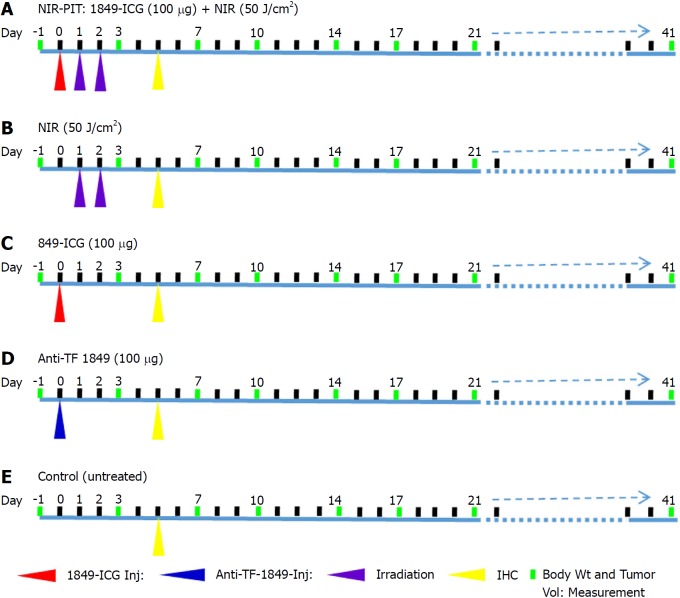
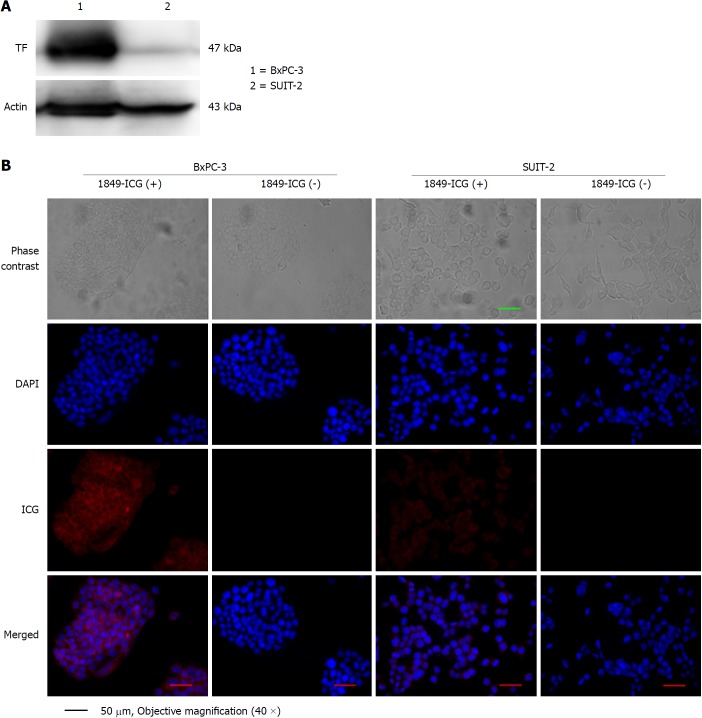
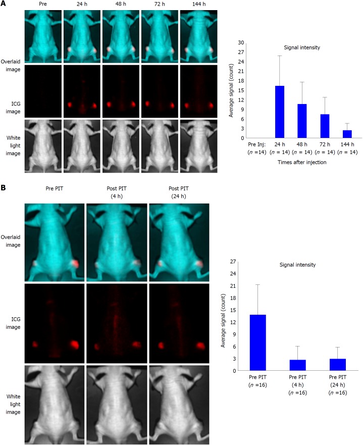
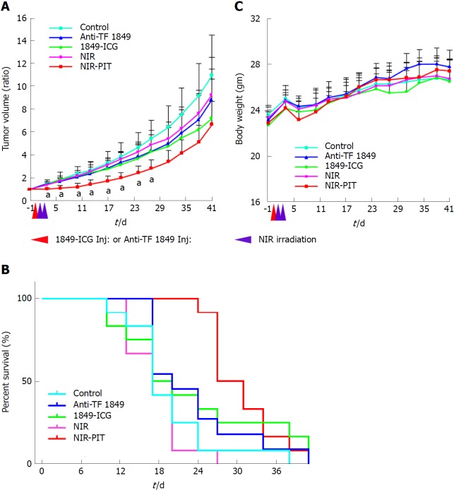
Near-infrared photoimmunotherapy (NIR-PIT) is a highly selective tumor treatment that utilizes an antibody-photosensitizer conjugate administration, followed by NIR light exposure. Anti-TF antibody 1849-ICG conjugate was synthesized by labeling of rat IgG anti-TF monoclonal antibody 1849 (anti-TF 1849) to a NIR photosensitizer, ICG. The expression levels of TF in two human pancreatic cancer cell lines were examined by western blotting. Specific binding of the 1849-ICG to TF-expressing BxPC-3 cells was examined by fluorescence microscopy. NIR-PIT-induced cell death was determined by cell viability imaging assay. longitudinal fluorescence imaging was used to explore the accumulation of 1849-ICG conjugate in xenograft tumors. To examine the effect of NIR-PIT, tumor-bearing mice were separated into 5 groups: (1) 100 μg of 1849-ICG i.v. administration followed by NIR light exposure (50 J/cm) on two consecutive days (Days 1 and 2); (2) NIR light exposure (50 J/cm) only on two consecutive days (Days 1 and 2); (3) 100 μg of 1849-ICG i.v. administration; (4) 100 μg of unlabeled anti-TF 1849 i.v. administration; and (5) the untreated control. Semiweekly tumor volume measurements, accompanied with histological and immunohistochemical (IHC) analyses of tumors, were performed 3 d after the 2 irradiation with NIR light to monitor the effect of treatments.
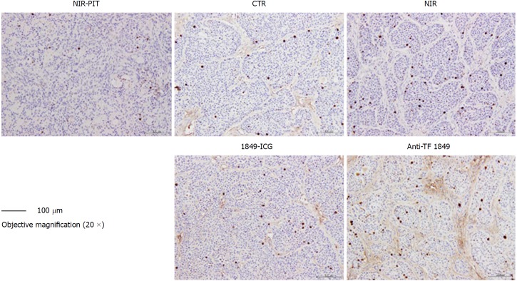
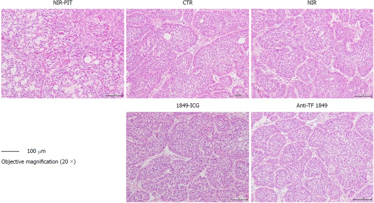
High TF expression in BxPC-3 cells was observed western blot analysis, concordant with the observed preferential binding with intracellular localization of 1849-ICG fluorescence microscopy. NIR-PIT-induced cell death was observed by performing cell viability imaging assay. In contrast to the other test groups, tumor growth was significantly inhibited by NIR-PIT with a statistically significant difference in relative tumor volumes for 27 d after the treatment start date [2.83 ± 0.38 (NIR-PIT) 5.42 ± 1.61 (Untreated), 4.90 ± 0.87 (NIR), 4.28 ± 1.87 (1849-ICG), 4.35 ± 1.42 (anti-TF 1849), at Day 27, < 0.05]. Tumors that received NIR-PIT showed evidence of necrotic cell death-associated features upon hematoxylin-eosin staining accompanied by a decrease in Ki-67-positive cells (a cell proliferation marker) by IHC examination.
The TF-targeted NIR-PIT with the 1849-ICG conjugate can potentially open a new platform for treatment of TF-expressing pancreatic cancer.
研究组织因子(TF)抗体与吲哚菁绿(ICG)偶联物介导的近红外光免疫治疗(NIR-PIT)在胰腺癌模型中的近红外光免疫治疗效果。
近红外光免疫治疗(NIR-PIT)是一种高度选择性的肿瘤治疗方法,利用抗体-光敏剂偶联物的给药,然后进行近红外光照射。通过将大鼠 IgG 抗 TF 单克隆抗体 1849(抗 TF 1849)标记到近红外光敏剂 ICG 上来合成抗 TF 抗体 1849-ICG 偶联物。通过蛋白质印迹分析检测两种人胰腺癌细胞系中 TF 的表达水平。通过荧光显微镜检查 1849-ICG 与 TF 表达的 BxPC-3 细胞的特异性结合。通过细胞活力成像测定法确定 NIR-PIT 诱导的细胞死亡。通过纵向荧光成像来探索 1849-ICG 偶联物在异种移植肿瘤中的积累。为了研究 NIR-PIT 的效果,将荷瘤小鼠分为 5 组:(1)静脉注射 100 μg 1849-ICG,然后在连续两天(第 1 天和第 2 天)进行近红外光照射(50 J/cm);(2)仅在连续两天(第 1 天和第 2 天)进行近红外光照射(50 J/cm);(3)静脉注射 100 μg 1849-ICG;(4)静脉注射 100 μg 未标记的抗 TF 1849;(5)未治疗的对照组。在两次近红外光照射后的第 3 天进行半周肿瘤体积测量,并进行肿瘤的组织学和免疫组织化学(IHC)分析,以监测治疗效果。
通过蛋白质印迹分析观察到 BxPC-3 细胞中 TF 的高表达,与通过荧光显微镜观察到的细胞内定位的 1849-ICG 的优先结合一致。通过进行细胞活力成像测定法观察到 NIR-PIT 诱导的细胞死亡。与其他实验组相比,NIR-PIT 显著抑制了肿瘤生长,在治疗开始后 27 天的相对肿瘤体积有统计学显著差异[2.83±0.38(NIR-PIT) 5.42±1.61(未治疗),4.90±0.87(NIR),4.28±1.87(1849-ICG),4.35±1.42(抗 TF 1849),在第 27 天,<0.05]。接受 NIR-PIT 的肿瘤在苏木精-伊红染色后显示出与坏死细胞死亡相关的特征性证据,并伴有 IHC 检查中 Ki-67 阳性细胞(细胞增殖标志物)的减少。
TF 靶向的 NIR-PIT 与 1849-ICG 偶联物可能为治疗 TF 表达的胰腺癌开辟新的平台。