Goldschleger Eye Institute, Sheba Medical Center, 52621, Tel-Hashomer, Israel.
Sackler Faculty of Medicine, Tel-Aviv University, 69978, Tel-Aviv, Israel.
J Nanobiotechnology. 2019 Jan 10;17(1):3. doi: 10.1186/s12951-018-0438-y.
Retinal degeneration diseases affect millions of patients worldwide and lead to incurable vision loss. These diseases are caused by pathologies in the retina and underlying choroid, located in the back of the eye. One of the major challenges in the development of treatments for these blinding diseases is the safe and efficient delivery of therapeutics into the back of the eye. Previous studies demonstrated that narrow size distribution core-shell near infra-red fluorescent iron oxide (IO) nanoparticles (NPs) coated with human serum albumin (HSA, IO/HSA NPs) increase the half-life of conjugated therapeutic factors, suggesting they may be used for sustained release of therapeutics. In the present study, the in vivo tracking by MRI and the long term safety of IO/HSA NPs delivery into the suprachoroid of a rat model of retinal degeneration were assessed.
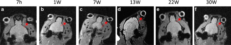
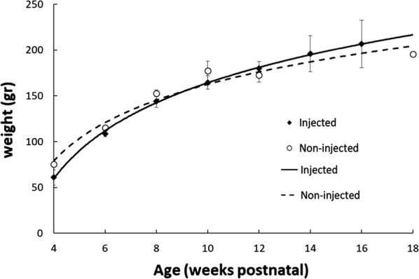
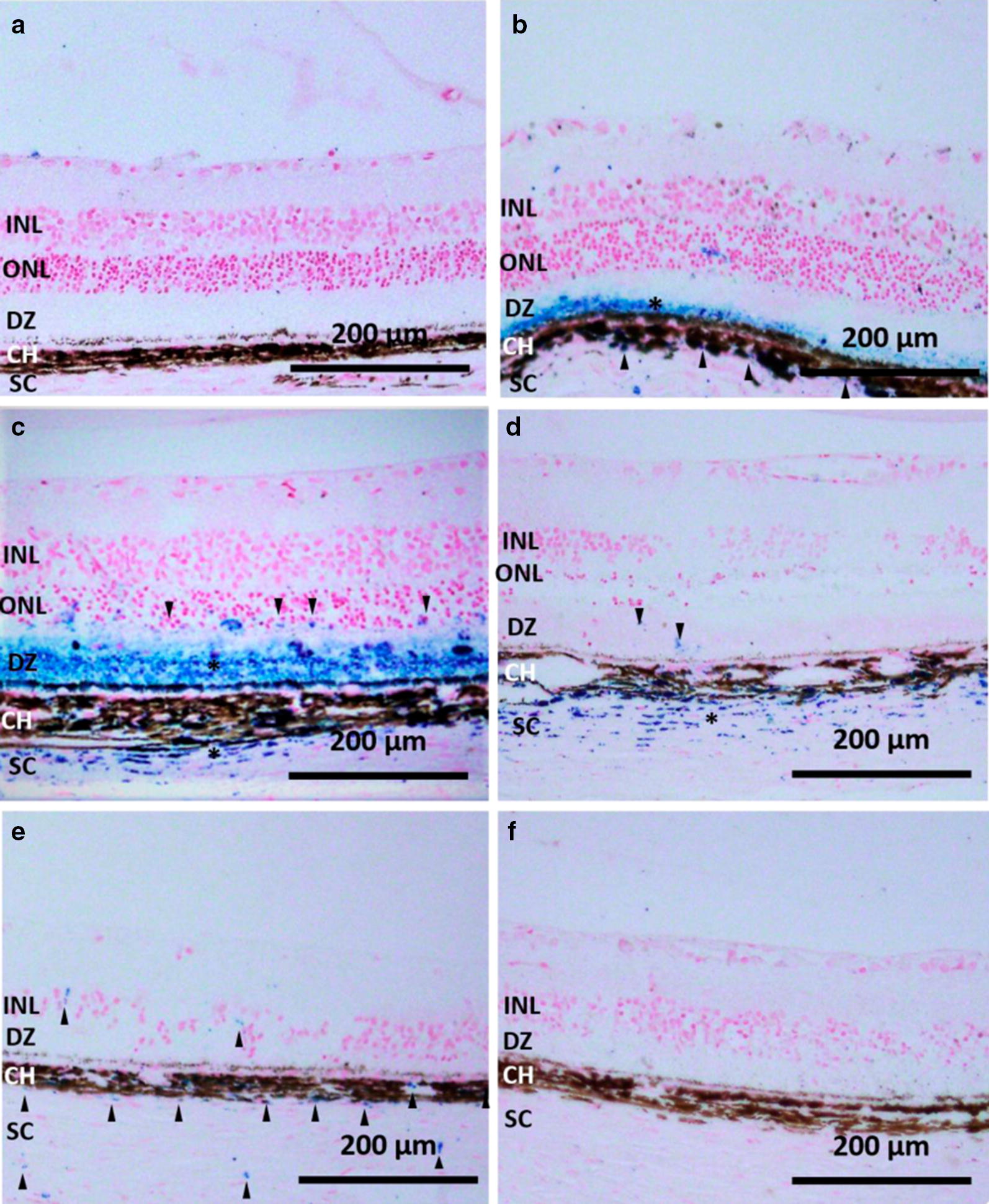
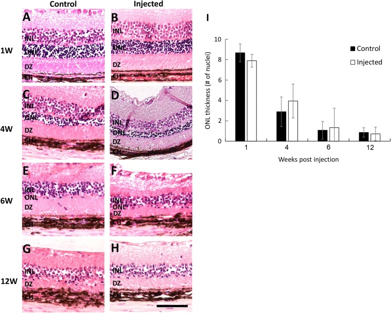
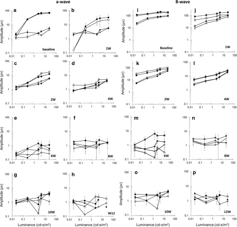
Twenty-five Royal College of Surgeons (RCS) pigmented rats received suprachoroidal injection of 20-nm IO/HSA NPs into the right eye. The left eye was not injected and used as control. Animals were examined by magnetic resonance imaging (MRI), electroretinogram (ERG) and histology up to 30 weeks following injection. IO/HSA NPs were detected in the back part of the rats' eyes up to 30 weeks following injection by MRI, and up to 6 weeks by histology. No significant differences in retinal structure and function were observed between injected and non-injected eyes. There was no significant difference in the weight of IO/HSA NP-injected animals compared to non-injected rats.
MRI could track the nanoparticles in the posterior segment of the injected eyes demonstrating their long-term persistence, and highlighting the possible use of MRI for translational studies in animals and in future clinical studies. Suprachoroidal injection of IO/HSA NPs showed no sign of adverse effects on retinal structure and function in a rat model of retinal degeneration, suggesting that suprachoroidal delivery of IO/HSA NPs is safe and that these NPs may be used in future translational and clinical studies for extended release drug delivery at the back of the eye.
视网膜退行性疾病影响着全球数以百万计的患者,导致不可治愈的视力丧失。这些疾病是由视网膜和位于眼睛后部的脉络膜的病理学引起的。在开发这些致盲疾病的治疗方法方面,面临的主要挑战之一是如何将治疗剂安全有效地递送到眼睛后部。先前的研究表明,用人血清白蛋白(HSA)包覆的窄粒径分布核壳近红外荧光氧化铁(IO)纳米颗粒(IO/HSA NPs)增加了共轭治疗因子的半衰期,表明它们可能用于治疗剂的持续释放。在本研究中,通过 MRI 对 IO/HSA NPs 递送至视网膜变性大鼠模型脉络膜上腔的体内示踪进行了评估,并对其长期安全性进行了研究。
25 只皇家外科学院(RCS)色素性大鼠右眼接受了 IO/HSA NPs 脉络膜上腔注射。左眼未注射,作为对照。动物在注射后 30 周内通过磁共振成像(MRI)、视网膜电图(ERG)和组织学检查进行评估。通过 MRI 可在注射后 30 周内检测到 IO/HSA NPs 在大鼠眼部的后部,通过组织学可在 6 周内检测到 IO/HSA NPs。注射眼和未注射眼的视网膜结构和功能无明显差异。与未注射大鼠相比,IO/HSA NP 注射动物的体重无明显差异。
MRI 可在注射眼的后节追踪纳米颗粒,证明其具有长期持久性,并突出了 MRI 在动物转化研究和未来临床研究中的潜在应用。IO/HSA NPs 脉络膜上腔注射在视网膜变性大鼠模型中未显示对视网膜结构和功能有不良影响,表明 IO/HSA NPs 脉络膜上腔给药是安全的,这些 NPs 可能在未来的转化和临床研究中用于在眼睛后部进行延长释放药物递送。