Katsuura Yoshihiro, Chang Eric, Sabri Shahbaaz A, Gardner Warren E, Doty Jesse F
Department of Orthopaedic Surgery, University of Tennessee College of Medicine Chattanooga, Chattanooga, TN (Dr. Katsuura, Dr. Sabri, Dr. Gardner, and Dr. Doty), and Tulane University School of Medicine, New Orleans, LA (Mr. Chang).
J Am Acad Orthop Surg Glob Res Rev. 2018 Aug 2;2(8):e034. doi: 10.5435/JAAOSGlobal-D-18-00034. eCollection 2018 Aug.
Multiple cadaver and radiographic analyses have been performed to define the surgical anatomy of the sacrum and pelvis. We provide a comprehensive review of this information, creating an accurate anatomic guide for practice and research.
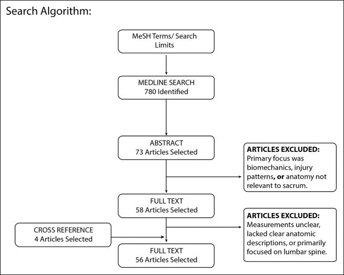
A systematic review was performed to identify publications citing sacral or iliac morphometric parameters based on cadaver or radiographic anatomy.
A total of 780 abstracts were evaluated. Fifty-six articles were included for final review and grouped into four sections: (1) bone density, (2) bony corridors, (3) screw length and trajectory, and (4) neurovascular and alimentary anatomy.
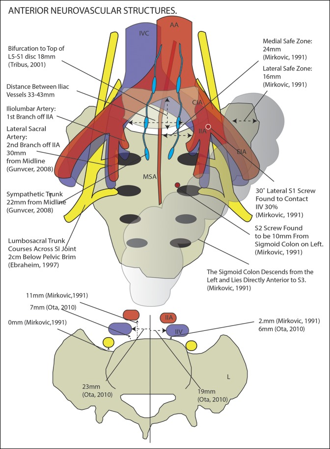
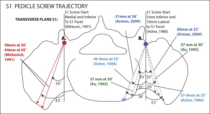
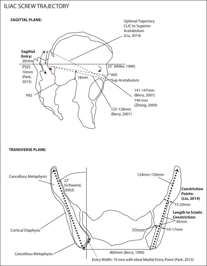
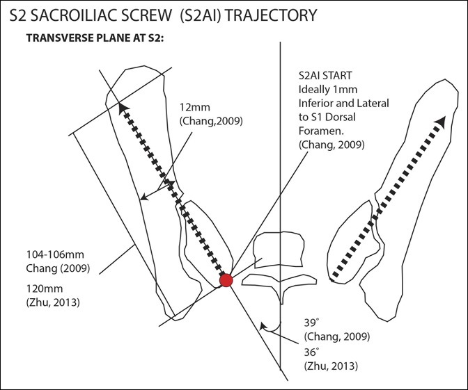
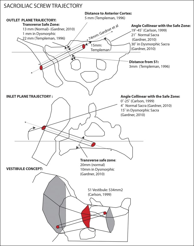
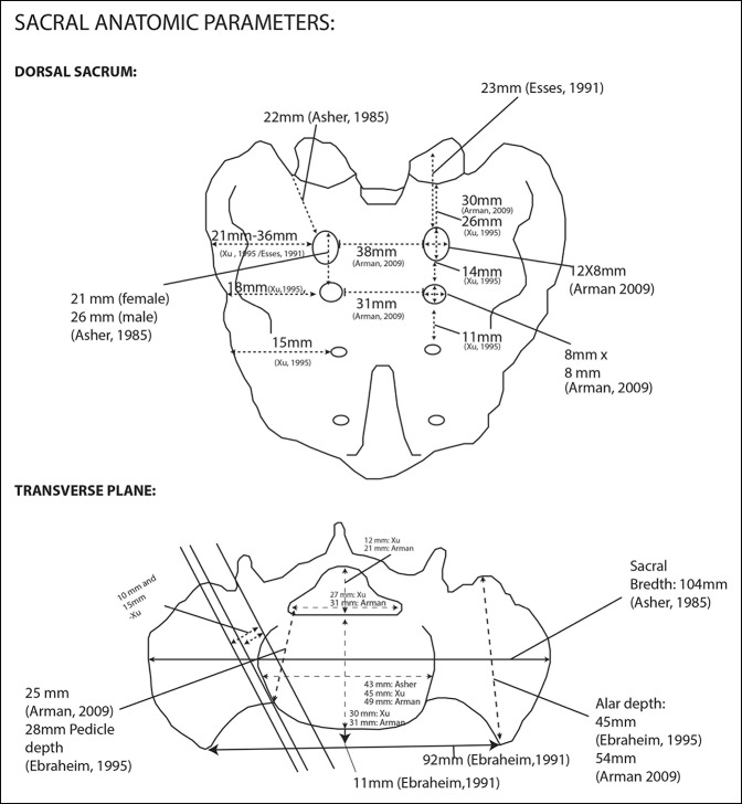
A systematic analysis of the radiographic and gross anatomic features of the sacrum has yet to be published. This review includes details on the spatial arrangement of the S1 and S2 pedicle screws, sacroiliac screws, iliac screws, S2 alar iliac screws, and pelvic neurovascular anatomy. The study can be referenced by clinicians for sacral dissection, implant application, and ongoing advances in orthopaedic research.
Systematic review.
Level IV.
已进行多项尸体和影像学分析以明确骶骨和骨盆的手术解剖结构。我们对这些信息进行全面综述,为临床实践和研究创建一份准确的解剖学指南。
进行系统综述,以识别基于尸体或影像学解剖引用骶骨或髂骨形态学参数的出版物。
共评估780篇摘要。56篇文章纳入最终综述,并分为四个部分:(1)骨密度,(2)骨通道,(3)螺钉长度和轨迹,以及(4)神经血管和消化道解剖结构。
关于骶骨影像学和大体解剖特征的系统分析尚未发表。本综述包括S1和S2椎弓根螺钉、骶髂螺钉、髂骨螺钉、S2翼状髂骨螺钉的空间排列以及骨盆神经血管解剖结构的详细信息。临床医生在进行骶骨解剖、植入物应用及骨科研究的持续进展时可参考本研究。
系统综述。
四级。