• 文献检索
  • 文档翻译
  • 深度研究
  • 学术资讯
  • Suppr Zotero 插件Zotero 插件
  • 邀请有礼
  • 套餐&价格
  • 历史记录
应用&插件
Suppr Zotero 插件Zotero 插件浏览器插件Mac 客户端Windows 客户端微信小程序
定价
高级版会员购买积分包购买API积分包
服务
文献检索文档翻译深度研究API 文档MCP 服务
关于我们
关于 Suppr公司介绍联系我们用户协议隐私条款
关注我们

Suppr 超能文献

核心技术专利:CN118964589B侵权必究
粤ICP备2023148730 号-1Suppr @ 2026

文献检索

告别复杂PubMed语法,用中文像聊天一样搜索,搜遍4000万医学文献。AI智能推荐,让科研检索更轻松。

立即免费搜索

文件翻译

保留排版,准确专业,支持PDF/Word/PPT等文件格式,支持 12+语言互译。

免费翻译文档

深度研究

AI帮你快速写综述,25分钟生成高质量综述,智能提取关键信息,辅助科研写作。

立即免费体验

用于骨科应用的含壳聚糖/镁钙硅酸盐复合材料的3D打印多孔钛合金支架的生物活性改善

Improved Bioactivity of 3D Printed Porous Titanium Alloy Scaffold with Chitosan/Magnesium-Calcium Silicate Composite for Orthopaedic Applications.

作者信息

Tsai Chun-Hao, Hung Chih-Hung, Kuo Che-Nan, Chen Cheng-Yu, Peng Yu-Ning, Shie Ming-You

机构信息

School of Medicine, China Medical University, Taichung 40447, Taiwan.

Department of Orthopedics, China Medical University Hospital, Taichung 40447, Taiwan.

出版信息

Materials (Basel). 2019 Jan 9;12(2):203. doi: 10.3390/ma12020203.

DOI:10.3390/ma12020203
PMID:30634440
原文链接:https://pmc.ncbi.nlm.nih.gov/articles/PMC6356721/
Abstract

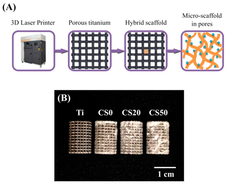
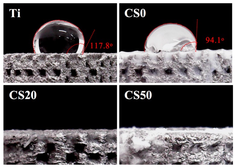
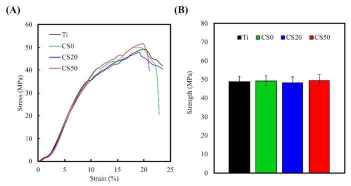
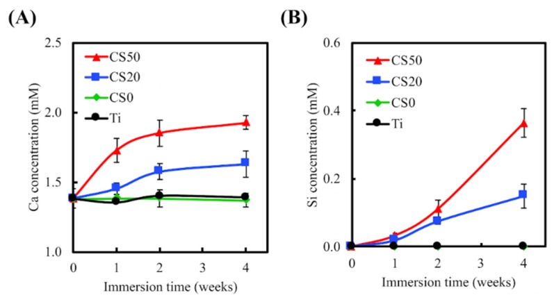
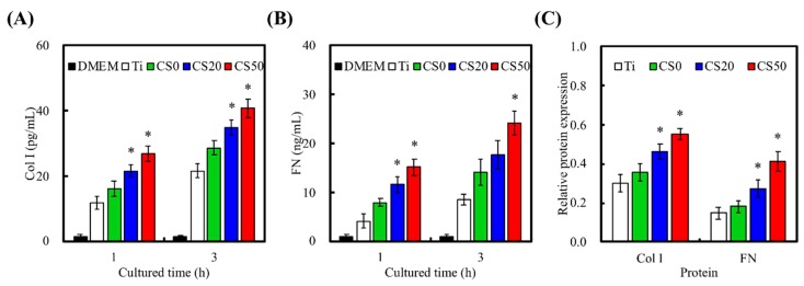
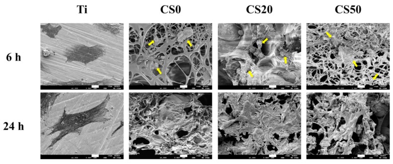
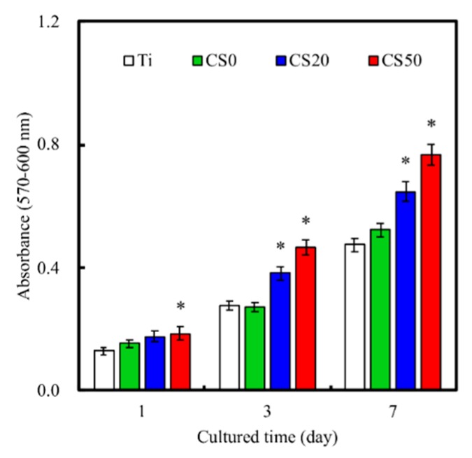
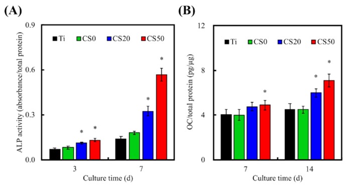
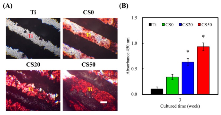
Recently, cases of bone defects have been increasing incrementally. Thus, repair or replacement of bone defects is gradually becoming a huge problem for orthopaedic surgeons. Three-dimensional (3D) scaffolds have since emerged as a potential candidate for bone replacement, of which titanium (Ti) alloys are one of the most promising candidates among the metal alloys due to their low cytotoxicity and mechanical properties. However, bioactivity remains a problem for metal alloys, which can be enhanced using simple immersion techniques to coat bioactive compounds onto the surface of Ti⁻6Al⁻4V scaffolds. In our study, we fabricated magnesium-calcium silicate (Mg⁻CS) and chitosan (CH) compounds onto Ti⁻6Al⁻4V scaffolds. Characterization of these surface-modified scaffolds involved an assessment of physicochemical properties as well as mechanical testing. Adhesion, proliferation, and growth of human Wharton's Jelly mesenchymal stem cells (WJMSCs) were assessed in vitro. In addition, the cell attachment morphology was examined using scanning electron microscopy to assess adhesion qualities. Osteogenic and mineralization assays were conducted to assess osteogenic expression. In conclusion, the Mg⁻CS/CH coated Ti⁻6Al⁻4V scaffolds were able to exhibit and retain pore sizes and their original morphologies and architectures, which significantly affected subsequent hard tissue regeneration. In addition, the surface was shown to be hydrophilic after modification and showed mechanical strength comparable to natural bone. Not only were our modified scaffolds able to match the mechanical properties of natural bone, it was also found that such modifications enhanced cellular behavior such as adhesion, proliferation, and differentiation, which led to enhanced osteogenesis and mineralization downstream. In vivo results indicated that Mg⁻CS/CH coated Ti⁻6Al⁻4V enhances the bone regeneration and ingrowth at the critical size bone defects of rabbits. These results indicated that the proposed Mg⁻CS/CH coated Ti⁻6Al⁻4V scaffolds exhibited a favorable, inducive micro-environment that could serve as a promising modification for future bone tissue engineering scaffolds.

摘要

近年来,骨缺损病例呈递增趋势。因此,骨缺损的修复或置换逐渐成为骨科医生面临的一大难题。三维(3D)支架应运而生,成为骨替代的潜在候选材料,其中钛(Ti)合金因其低细胞毒性和机械性能,成为金属合金中最有前景的候选材料之一。然而,生物活性仍是金属合金面临的一个问题,可以通过简单的浸泡技术将生物活性化合物涂覆在Ti⁻6Al⁻4V支架表面来增强其生物活性。在我们的研究中,我们在Ti⁻6Al⁻4V支架上制备了镁钙硅酸盐(Mg⁻CS)和壳聚糖(CH)化合物。对这些表面改性支架的表征包括物理化学性质评估以及力学测试。在体外评估了人脐带华通氏胶间充质干细胞(WJMSCs)的黏附、增殖和生长情况。此外,使用扫描电子显微镜检查细胞附着形态以评估黏附质量。进行了成骨和矿化测定以评估成骨表达。总之,Mg⁻CS/CH涂层的Ti⁻6Al⁻4V支架能够展现并保持孔径及其原始形态和结构,这对随后的硬组织再生有显著影响。此外,改性后的表面显示出亲水性,并具有与天然骨相当的机械强度。我们的改性支架不仅能够匹配天然骨的机械性能,还发现这种改性增强了细胞行为,如黏附、增殖和分化,从而导致下游成骨和矿化增强。体内结果表明,Mg⁻CS/CH涂层的Ti⁻6Al⁻4V可促进兔临界尺寸骨缺损处的骨再生和骨长入。这些结果表明,所提出的Mg⁻CS/CH涂层的Ti⁻6Al⁻4V支架展现出良好的、诱导性的微环境,有望成为未来骨组织工程支架的一种有前景的改性材料。

https://cdn.ncbi.nlm.nih.gov/pmc/blobs/d3af/6356721/145bd0b62c89/materials-12-00203-g012.jpg
https://cdn.ncbi.nlm.nih.gov/pmc/blobs/d3af/6356721/270f60aad10a/materials-12-00203-g001.jpg
https://cdn.ncbi.nlm.nih.gov/pmc/blobs/d3af/6356721/9d7d7ce0fb15/materials-12-00203-g002.jpg
https://cdn.ncbi.nlm.nih.gov/pmc/blobs/d3af/6356721/c3a838ea8807/materials-12-00203-g003.jpg
https://cdn.ncbi.nlm.nih.gov/pmc/blobs/d3af/6356721/bc774ab4df50/materials-12-00203-g004.jpg
https://cdn.ncbi.nlm.nih.gov/pmc/blobs/d3af/6356721/5f9f3d93538a/materials-12-00203-g005.jpg
https://cdn.ncbi.nlm.nih.gov/pmc/blobs/d3af/6356721/3533e64cf150/materials-12-00203-g006.jpg
https://cdn.ncbi.nlm.nih.gov/pmc/blobs/d3af/6356721/325d67dbc725/materials-12-00203-g007.jpg
https://cdn.ncbi.nlm.nih.gov/pmc/blobs/d3af/6356721/6f6aaf119ff3/materials-12-00203-g008.jpg
https://cdn.ncbi.nlm.nih.gov/pmc/blobs/d3af/6356721/ef47a66d3072/materials-12-00203-g009.jpg
https://cdn.ncbi.nlm.nih.gov/pmc/blobs/d3af/6356721/784c1dc58744/materials-12-00203-g010.jpg
https://cdn.ncbi.nlm.nih.gov/pmc/blobs/d3af/6356721/2fe239dc6df7/materials-12-00203-g011.jpg
https://cdn.ncbi.nlm.nih.gov/pmc/blobs/d3af/6356721/145bd0b62c89/materials-12-00203-g012.jpg

相似文献

1
Improved Bioactivity of 3D Printed Porous Titanium Alloy Scaffold with Chitosan/Magnesium-Calcium Silicate Composite for Orthopaedic Applications.用于骨科应用的含壳聚糖/镁钙硅酸盐复合材料的3D打印多孔钛合金支架的生物活性改善
Materials (Basel). 2019 Jan 9;12(2):203. doi: 10.3390/ma12020203.
2
Combined Effects of Polydopamine-Assisted Copper Immobilization on 3D-Printed Porous Ti6Al4V Scaffold for Angiogenic and Osteogenic Bone Regeneration.多巴胺辅助铜固定化对用于血管生成和成骨骨再生的 3D 打印多孔 Ti6Al4V 支架的联合效应。
Cells. 2022 Sep 9;11(18):2824. doi: 10.3390/cells11182824.
3
Bioactive calcium silicate/poly-ε-caprolactone composite scaffolds 3D printed under mild conditions for bone tissue engineering.在温和条件下 3D 打印的生物活性硅酸钙/聚己内酯复合支架用于骨组织工程。
J Mater Sci Mater Med. 2017 Dec 27;29(1):11. doi: 10.1007/s10856-017-6020-6.
4
Adhesion, proliferation, and osteogenic differentiation of human mesenchymal stem cells on additively manufactured Ti6Al4V alloy scaffolds modified with calcium phosphate nanoparticles.钙磷纳米粒子改性增材制造 Ti6Al4V 合金支架上的人骨髓间充质干细胞的黏附、增殖和成骨分化。
Colloids Surf B Biointerfaces. 2019 Apr 1;176:130-139. doi: 10.1016/j.colsurfb.2018.12.047. Epub 2018 Dec 18.
5
Large-pore-size Ti6Al4V scaffolds with different pore structures for vascularized bone regeneration.大孔径 Ti6Al4V 支架具有不同的孔隙结构,可用于血管化骨再生。
Mater Sci Eng C Mater Biol Appl. 2021 Dec;131:112499. doi: 10.1016/j.msec.2021.112499. Epub 2021 Oct 19.
6
A pH-neutral bioactive glass coated 3D-printed porous Ti6Al4V scaffold with enhanced osseointegration.一种 pH 值中性的生物活性玻璃涂层 3D 打印多孔 Ti6Al4V 支架,具有增强的骨整合能力。
J Mater Chem B. 2023 Feb 8;11(6):1203-1212. doi: 10.1039/d2tb02129c.
7
Osteogenesis of 3D printed porous Ti6Al4V implants with different pore sizes.3D 打印不同孔径多孔 Ti6Al4V 植入物的成骨作用。
J Mech Behav Biomed Mater. 2018 Aug;84:1-11. doi: 10.1016/j.jmbbm.2018.04.010. Epub 2018 Apr 18.
8
Additive manufacturing of barium-doped calcium silicate/poly-ε-caprolactone scaffolds to activate CaSR and AKT signalling and osteogenic differentiation of mesenchymal stem cells.钡掺杂硅酸钙/聚己内酯支架的增材制造激活钙敏感受体和 AKT 信号通路并促进间充质干细胞的成骨分化。
J Mater Chem B. 2023 May 31;11(21):4666-4676. doi: 10.1039/d3tb00208j.
9
The synergistic effects of graphene-contained 3D-printed calcium silicate/poly-ε-caprolactone scaffolds promote FGFR-induced osteogenic/angiogenic differentiation of mesenchymal stem cells.含石墨烯的 3D 打印硅酸钙/聚己内酯支架的协同作用促进成纤维细胞生长因子受体诱导的间充质干细胞成骨/血管生成分化。
Mater Sci Eng C Mater Biol Appl. 2019 Nov;104:109887. doi: 10.1016/j.msec.2019.109887. Epub 2019 Jun 24.
10
3D porous Ti6Al4V-beta-tricalcium phosphate scaffolds directly fabricated by additive manufacturing.3D 多孔 Ti6Al4V-β-磷酸三钙支架的直接增材制造。
Acta Biomater. 2021 May;126:496-510. doi: 10.1016/j.actbio.2021.03.021. Epub 2021 Mar 13.

引用本文的文献

1
3D Printing and Surface Engineering of Ti6Al4V Scaffolds for Enhanced Osseointegration in an In Vitro Study.用于体外研究中增强骨整合的Ti6Al4V支架的3D打印与表面工程
Biomimetics (Basel). 2024 Jul 10;9(7):423. doi: 10.3390/biomimetics9070423.
2
Recent Advancements in Bone Tissue Engineering: Integrating Smart Scaffold Technologies and Bio-Responsive Systems for Enhanced Regeneration.骨组织工程的最新进展:智能支架技术与生物响应系统的整合,以增强再生。
Int J Mol Sci. 2024 May 30;25(11):6012. doi: 10.3390/ijms25116012.
3
Additively manufactured porous scaffolds by design for treatment of bone defects.

本文引用的文献

1
Electrospun multicomponent and multifunctional nanofibrous bone tissue engineering scaffolds.静电纺多组分多功能纳米纤维骨组织工程支架
J Mater Chem B. 2017 Feb 21;5(7):1388-1399. doi: 10.1039/c6tb02907h. Epub 2017 Jan 27.
2
Silicate-based bioceramics regulating osteoblast differentiation through a BMP2 signalling pathway.基于硅酸盐的生物陶瓷通过骨形态发生蛋白2信号通路调节成骨细胞分化。
J Mater Chem B. 2017 Sep 21;5(35):7297-7306. doi: 10.1039/c7tb01931a. Epub 2017 Aug 23.
3
Effects of magnesium silicate on the mechanical properties, biocompatibility, bioactivity, degradability, and osteogenesis of poly(butylene succinate)-based composite scaffolds for bone repair.
通过设计增材制造的多孔支架用于治疗骨缺损。
Front Bioeng Biotechnol. 2024 Jan 19;11:1252636. doi: 10.3389/fbioe.2023.1252636. eCollection 2023.
4
The Importance of Chitosan Coatings in Dentistry.壳聚糖涂层在牙科中的重要性。
Mar Drugs. 2023 Nov 26;21(12):613. doi: 10.3390/md21120613.
5
A surface metal ion-modified 3D-printed Ti-6Al-4V implant with direct and immunoregulatory antibacterial and osteogenic activity.一种具有直接和免疫调节抗菌及成骨活性的表面金属离子改性3D打印Ti-6Al-4V植入物。
Front Bioeng Biotechnol. 2023 Mar 16;11:1142264. doi: 10.3389/fbioe.2023.1142264. eCollection 2023.
6
Cell Adhesion and Initial Bone Matrix Deposition on Titanium-Based Implants with Chitosan-Collagen Coatings: An In Vitro Study.壳聚糖-胶原蛋白涂层钛基种植体表面细胞黏附及初始骨基质沉积的体外研究。
Int J Mol Sci. 2023 Mar 2;24(5):4810. doi: 10.3390/ijms24054810.
7
Three-Dimensional Impression of Biomaterials for Alveolar Graft: Scoping Review.用于牙槽嵴植骨的生物材料的三维印记:范围综述
J Funct Biomater. 2023 Jan 29;14(2):76. doi: 10.3390/jfb14020076.
8
Synthetic materials in craniofacial regenerative medicine: A comprehensive overview.颅面再生医学中的合成材料:全面综述。
Front Bioeng Biotechnol. 2022 Nov 9;10:987195. doi: 10.3389/fbioe.2022.987195. eCollection 2022.
9
Combined Effects of Polydopamine-Assisted Copper Immobilization on 3D-Printed Porous Ti6Al4V Scaffold for Angiogenic and Osteogenic Bone Regeneration.多巴胺辅助铜固定化对用于血管生成和成骨骨再生的 3D 打印多孔 Ti6Al4V 支架的联合效应。
Cells. 2022 Sep 9;11(18):2824. doi: 10.3390/cells11182824.
10
Advanced Surface Modification for 3D-Printed Titanium Alloy Implant Interface Functionalization.用于3D打印钛合金植入物界面功能化的先进表面改性
Front Bioeng Biotechnol. 2022 Mar 1;10:850110. doi: 10.3389/fbioe.2022.850110. eCollection 2022.
硅酸镁对用于骨修复的聚丁二酸丁二醇酯基复合支架的力学性能、生物相容性、生物活性、降解性和成骨作用的影响。
J Mater Chem B. 2016 Dec 28;4(48):7974-7988. doi: 10.1039/c6tb02429g. Epub 2016 Nov 25.
4
3D plotting of highly uniform Sr(PO)SiO bioceramic scaffolds for bone tissue engineering.用于骨组织工程的高度均匀的Sr(PO)SiO生物陶瓷支架的3D绘图。
J Mater Chem B. 2016 Oct 7;4(37):6200-6212. doi: 10.1039/c6tb01692h. Epub 2016 Sep 7.
5
Enhanced adhesion and differentiation of human mesenchymal stem cell inside apatite-mineralized/poly(dopamine)-coated poly(ε-caprolactone) scaffolds by stereolithography.通过立体光刻技术增强人骨髓间充质干细胞在磷灰石矿化/聚多巴胺涂层聚己内酯支架内的黏附与分化。
J Mater Chem B. 2016 Oct 14;4(38):6307-6315. doi: 10.1039/c6tb01377e. Epub 2016 Sep 12.
6
Stimulatory effects of the fast setting and suitable degrading Ca-Si-Mg cement on both cementogenesis and angiogenesis differentiation of human periodontal ligament cells.快凝且降解适宜的钙硅镁水泥对人牙周膜细胞牙骨质生成和血管生成分化的刺激作用
J Mater Chem B. 2015 Sep 21;3(35):7099-7108. doi: 10.1039/c5tb00713e. Epub 2015 Aug 14.
7
Containerless processing for preparation of akermanite bioceramic spheres with homogeneous structure, tailored bioactivity and degradation.用于制备具有均匀结构、定制生物活性和降解性能的钙黄长石生物陶瓷球的无容器加工。
J Mater Chem B. 2013 Feb 21;1(7):1019-1026. doi: 10.1039/c2tb00215a. Epub 2012 Dec 17.
8
Accelerated host angiogenesis and immune responses by ion release from mesoporous bioactive glass.介孔生物活性玻璃离子释放促进宿主血管生成和免疫反应。
J Mater Chem B. 2018 May 28;6(20):3274-3284. doi: 10.1039/c8tb00683k. Epub 2018 May 3.
9
The Physicochemical Properties of Decellularized Extracellular Matrix-Coated 3D Printed Poly(ε-caprolactone) Nerve Conduits for Promoting Schwann Cells Proliferation and Differentiation.用于促进雪旺细胞增殖和分化的去细胞细胞外基质涂层3D打印聚(ε-己内酯)神经导管的物理化学性质
Materials (Basel). 2018 Sep 9;11(9):1665. doi: 10.3390/ma11091665.
10
Surface Modification of Calcium Silicate via Mussel-Inspired Polydopamine and Effective Adsorption of Extracellular Matrix to Promote Osteogenesis Differentiation for Bone Tissue Engineering.通过贻贝启发的聚多巴胺对硅酸钙进行表面改性及细胞外基质的有效吸附以促进骨组织工程中的成骨分化
Materials (Basel). 2018 Sep 9;11(9):1664. doi: 10.3390/ma11091664.