Department of Radiation Sciences, Umeå University, Umeå, Sweden.
Umeå Center for Functional Brain Imaging, Umeå University, Umeå, Sweden.
J Magn Reson Imaging. 2019 Aug;50(2):511-518. doi: 10.1002/jmri.26641. Epub 2019 Jan 14.
Accelerated 4D flow MRI allows for high-resolution velocity measurements with whole-brain coverage. Such scans are increasingly used to calculate flow rates of individual arteries in the vascular tree, but detailed information about the accuracy and precision in relation to different postprocessing options is lacking.
To evaluate and optimize three proposed segmentation methods and determine the accuracy of in vivo 4D flow MRI blood flow rate assessments in major cerebral arteries, with high-resolution 2D PCMRI as a reference.
Prospective.
Thirty-five subjects (20 women, 79 ± 5 years, range 70-91 years).
FIELD STRENGTH/SEQUENCE: 4D flow MRI with PC-VIPR and 2D PCMRI acquired with a 3 T scanner.
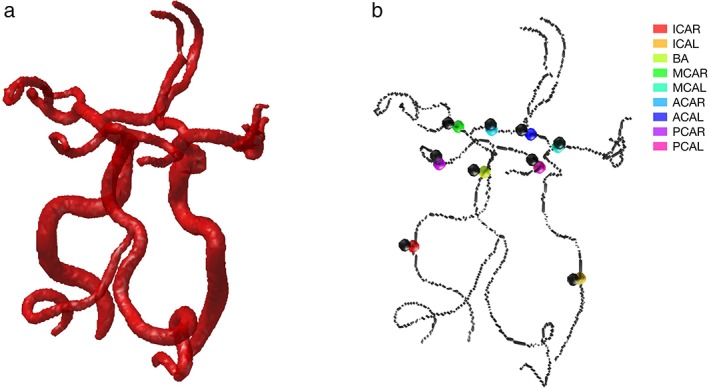
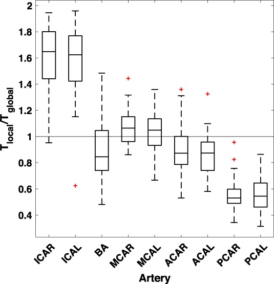
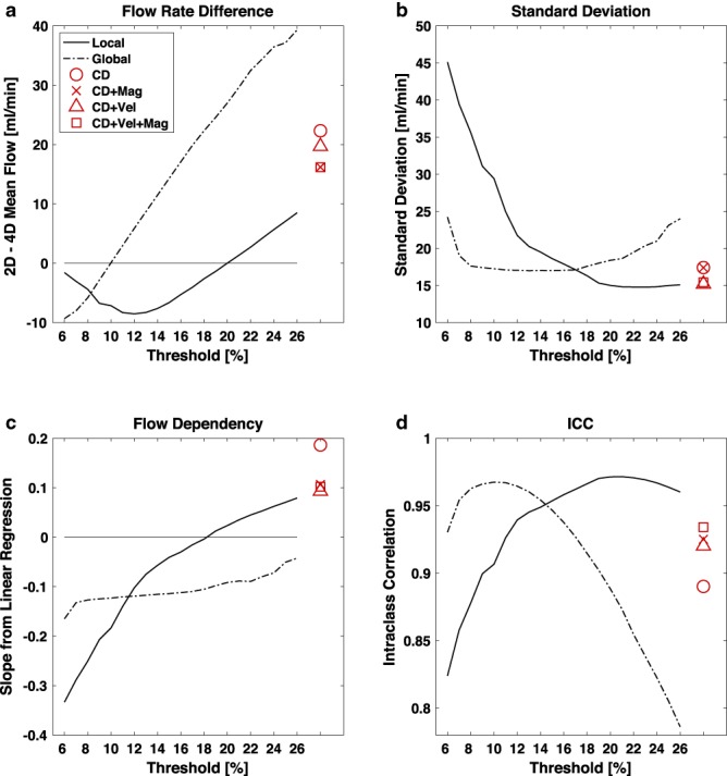
We compared blood flow rates measured with 4D flow MRI, to the reference, in nine main cerebral arteries. Lumen segmentation in the 4D flow MRI was performed with k-means clustering using four different input datasets, and with two types of thresholding methods. The threshold was defined as a percentage of the maximum intensity value in the complex difference image. Local and global thresholding approaches were used, with evaluated thresholds from 6-26%.
Paired t-test, F-test, linear correlation (P < 0.05 was considered significant) along with intraclass correlation (ICC).
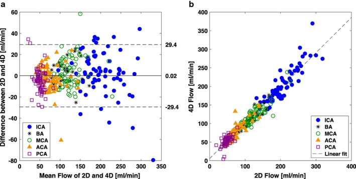
With the thresholding methods, the lowest average flow difference was obtained for 20% local (0.02 ± 15.0 ml/min, ICC = 0.97, n = 310) or 10% global (0.08 ± 17.3 ml/min, ICC = 0.97, n = 310) thresholding with a significant lower standard deviation for local (F-test, P = 0.01). For all clustering methods, we found a large systematic underestimation of flow compared with 2D PCMRI (16.1-22.3 ml/min).
A locally adapted threshold value gives a more stable result compared with a globally fixed threshold. 4D flow with the proposed segmentation method has the potential to become a useful reliable clinical tool for assessment of blood flow in the major cerebral arteries.
2 Technical Efficacy: Stage 2 J. Magn. Reson. Imaging 2019;50:511-518.
加速 4D 流 MRI 可实现具有全脑覆盖的高分辨率速度测量。此类扫描越来越多地用于计算血管树中各个动脉的流量,但关于不同后处理选项的准确性和精密度的详细信息却很缺乏。
评估和优化三种拟议的分割方法,并确定高分辨率 2D PCMRI 作为参考时,体内 4D 流 MRI 血流率评估在主要脑动脉中的准确性。
前瞻性。
35 名受试者(20 名女性,79 ± 5 岁,范围 70-91 岁)。
磁场强度/序列:使用 PC-VIPR 进行 4D 流 MRI 采集,在 3T 扫描仪上进行 2D PCMRI 采集。
我们将 4D 流 MRI 测量的血流率与参考值进行了比较,比较了 9 条主要脑动脉中的血流率。4D 流 MRI 中的管腔分割使用 k-均值聚类方法,使用四种不同的输入数据集和两种类型的阈值方法进行。阈值定义为复差异图像中最大强度值的百分比。使用局部和全局阈值方法,评估的阈值范围为 6-26%。
配对 t 检验、F 检验、线性相关(P < 0.05 为有统计学意义)以及组内相关(ICC)。
使用阈值方法,局部 20%(0.02 ± 15.0 ml/min,ICC = 0.97,n = 310)或全局 10%(0.08 ± 17.3 ml/min,ICC = 0.97,n = 310)的阈值可获得最低的平均流量差异,局部的标准偏差显著降低(F 检验,P = 0.01)。对于所有聚类方法,与 2D PCMRI 相比,我们发现血流存在较大的系统低估(16.1-22.3 ml/min)。
与全局固定阈值相比,局部自适应阈值可获得更稳定的结果。拟议分割方法的 4D 流具有成为评估主要脑动脉血流的有用可靠临床工具的潜力。
2 技术功效:第 2 阶段 J. Magn. Reson. Imaging 2019;50:511-518.