Division of Rheumatology, Allergy and Immunology and the Thurston Arthritis Research Center, University of North Carolina at Chapel Hill, Chapel Hill, NC, USA.
Division of Rheumatology, Allergy and Immunology and the Thurston Arthritis Research Center, University of North Carolina at Chapel Hill, Chapel Hill, NC, USA; Nanoscience and Nanoengineering, South Dakota School of Mines and Technology, BioSNTR, Rapid City, SD, USA.
Free Radic Biol Med. 2019 Apr;134:139-152. doi: 10.1016/j.freeradbiomed.2019.01.005. Epub 2019 Jan 9.
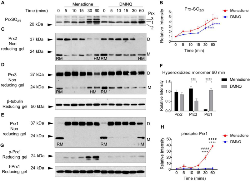
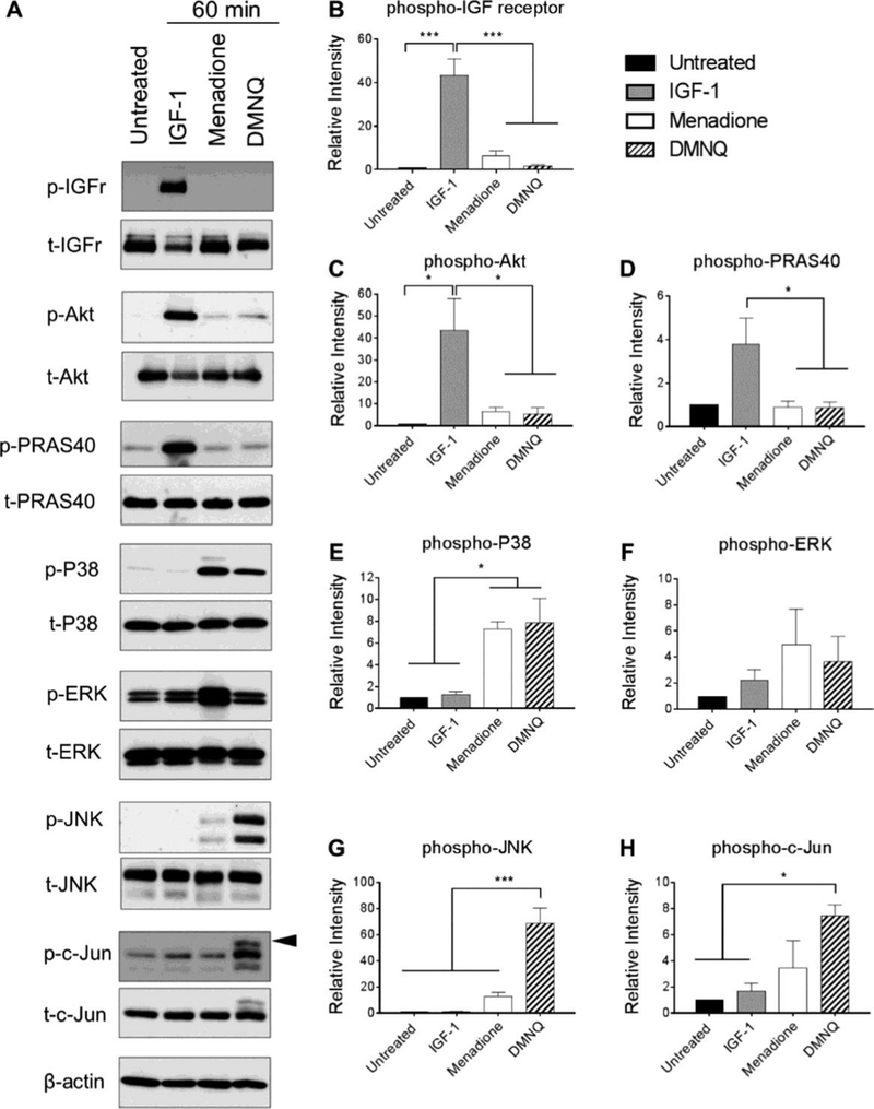
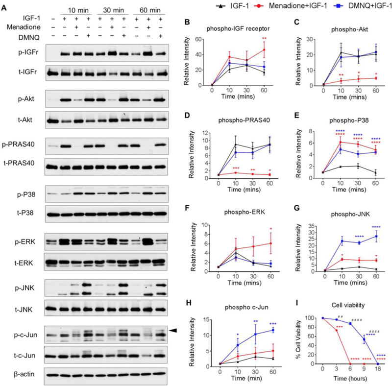
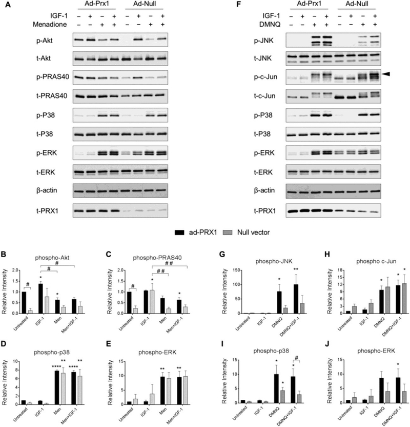
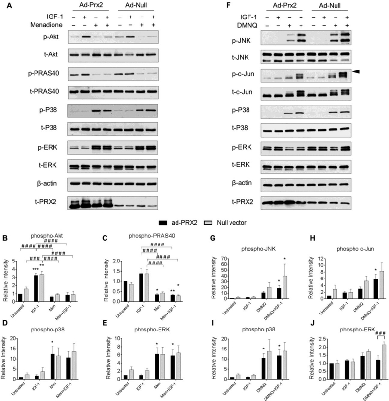
The peroxiredoxin (Prx) family of Cys-dependent peroxidases control intracellular levels of HO and can regulate signal transduction. Inhibition of the Prxs, through hyperoxidation amongst other mechanisms, leads to oxidative stress conditions that can alter homeostatic signaling. To determine the effects oxidation of Prx1-Prx3 has on MAP kinase and IGF-1 signaling events in human chondrocytes, this study used 2-methyl-1,4-naphthoquinone (menadione) and 2,3-dimethyl-1,4-naphthoquinone (DMNQ) as HO-generating tools due to their differential mechanisms of action. Menadione and DMNQ generated similar levels of intracellular HO as determined using the biosensor Orp1-roGFP and by measuring Prx redox status. However, menadione generated higher levels of mitochondrial HO associated with Prx3 hyperoxidation and phosphorylation of Prx1 while DMNQ treatment was associated with hyperoxidation of cytosolic Prx1 and Prx2 but not mitochondrial Prx3. Both menadione and DMNQ induced sustained phosphorylation of p38 but only DMNQ activated JNK. Menadione but not DMNQ inhibited IGF-1-induced Akt phosphorylation. Chondrocytes transduced with an adenoviral vector to overexpress Prx3 displayed decreased PrxSO formation in response to menadione which was associated with restoration of IGF-1-mediated Akt signaling and inhibition of p38 phosphorylation. Prx1 and Prx2 overexpression had no effects on Prx redox status but Prx1 overexpression enhanced basal Akt phosphorylation. These results suggest that hyperoxidation of specific Prx isoforms is associated with distinct cell signaling events and identify Prx3 redox status as an important regulator of anabolic and catabolic signal transduction. Targeted strategies to prevent mitochondrial Prx3 hyperoxidation could be useful in maintaining cellular redox balance and homeostatic signaling.
过氧化物酶(Prx)家族的 Cys 依赖性过氧化物酶控制 HO 的细胞内水平,并可以调节信号转导。通过过氧化物酶的超氧化等机制抑制过氧化物酶,会导致氧化应激条件,从而改变体内平衡信号。为了确定 Prx1-Prx3 的氧化对人软骨细胞中 MAP 激酶和 IGF-1 信号事件的影响,本研究使用 2-甲基-1,4-萘醌(甲萘醌)和 2,3-二甲基-1,4-萘醌(DMNQ)作为 HO 生成工具,因为它们的作用机制不同。甲萘醌和 DMNQ 产生类似水平的细胞内 HO,如使用生物传感器 Orp1-roGFP 确定的和通过测量 Prx 氧化还原状态确定的。然而,甲萘醌生成与 Prx3 过氧化物酶和磷酸化相关的更高水平的线粒体 HO,而 DMNQ 处理与细胞质 Prx1 和 Prx2 的过氧化物酶,但不是线粒体 Prx3 的过氧化物酶相关。甲萘醌和 DMNQ 均诱导 p38 的持续磷酸化,但只有 DMNQ 激活 JNK。甲萘醌但不是 DMNQ 抑制 IGF-1 诱导的 Akt 磷酸化。用腺病毒载体过表达 Prx3 的软骨细胞显示出对甲萘醌的反应性 PrxSO 形成减少,这与 IGF-1 介导的 Akt 信号的恢复和 p38 磷酸化的抑制有关。Prx1 和 Prx2 的过表达对 Prx 氧化还原状态没有影响,但 Prx1 的过表达增强了基础 Akt 磷酸化。这些结果表明,特定 Prx 同工型的过氧化物酶与不同的细胞信号事件相关,并且确定 Prx3 的氧化还原状态是合成代谢和分解代谢信号转导的重要调节剂。防止线粒体 Prx3 过氧化物酶的靶向策略可能有助于维持细胞氧化还原平衡和体内平衡信号。