Department of Clinical Biochemistry, Aalborg University Hospital, Aalborg, Denmark.
Department of Clinical Medicine, Aalborg University, Aalborg, Denmark.
PLoS One. 2019 Jan 14;14(1):e0210835. doi: 10.1371/journal.pone.0210835. eCollection 2019.
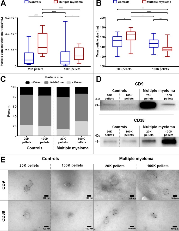
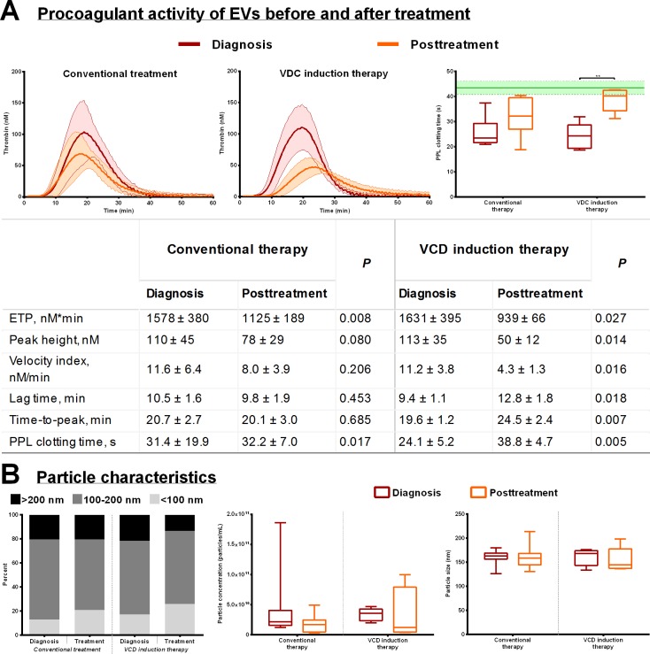
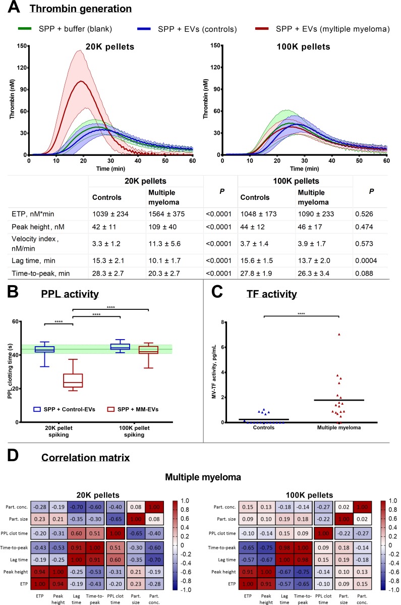
Multiple myeloma (MM) patients have increased risk of developing venous thromboembolism, but the underlying mechanisms and the effect on the coagulation system of the disease and the current cancer therapies are not known. It is possible that cancer-associated extracellular vesicles (EV), carrying tissue factor (TF) and procoagulant phospholipids (PPL) may play a role in thrombogenesis. The aim of this study was to perform an in-depth analysis of procoagulant activity of small and large EVs isolated from 20 MM patients at diagnosis and after receiving first-line treatment compared with 20 healthy control subjects. Differential ultracentrifugation at 20,000 × g and 100,000 × g were used to isolate EVs for quantitative and phenotypical analysis through nanoparticle tracking analysis, Western blotting and transmission electron microscopy. The isolated EVs were analyzed for procoagulant activity using the calibrated automated thrombogram technique, a factor Xa-based activity assay, and the STA Procoag-PPL assay. In general, MM patients contained more EVs, and immunoelectron microscopy confirmed the presence of CD9- and CD38-positive EVs. EVs in the 20,000 × g pellets from MM patients exerted procoagulant activity visualized by increased thrombin generation and both TF and PPL activity. This effect diminished during treatment, with the most prominent effect observed in the high-dose chemotherapy eligible patients after induction therapy with bortezomib, cyclophosphamide, and dexamethasone. In conclusion, the EVs in patients with MM carrying TF and PPL are thus capable of exerting procoagulant activity.
多发性骨髓瘤(MM)患者发生静脉血栓栓塞的风险增加,但疾病和当前癌症治疗对凝血系统的潜在机制和影响尚不清楚。携带组织因子(TF)和促凝磷脂(PPL)的癌相关细胞外囊泡(EV)可能在血栓形成中起作用。本研究旨在深入分析 20 名 MM 患者在诊断时和接受一线治疗后与 20 名健康对照者分离的小和大 EV 的促凝活性。通过纳米颗粒跟踪分析、Western blot 和透射电子显微镜,使用 20,000×g 和 100,000×g 进行差速超速离心来分离 EV,用于定量和表型分析。使用校准的自动化血栓图技术、基于因子 Xa 的活性测定和 STA Procoag-PPL 测定分析分离的 EV 的促凝活性。一般来说,MM 患者含有更多的 EV,免疫电子显微镜证实存在 CD9-和 CD38-阳性 EV。MM 患者 20,000×g 沉淀物中的 EV 表现出促凝活性,表现为凝血酶生成增加以及 TF 和 PPL 活性增加。这种作用在治疗期间减弱,在接受硼替佐米、环磷酰胺和地塞米松诱导治疗的高剂量化疗合格患者中观察到最明显的作用。总之,MM 患者携带 TF 和 PPL 的 EV 因此能够发挥促凝活性。