Liu Chao, Cabahug-Zuckerman Pamela, Stubbs Christopher, Pendola Martin, Cai Cinyee, Mann Kenneth A, Castillo Alesha B
Department of Biomedical Engineering, Tandon School of Engineering, New York University, New York, NY, USA.
Department of Orthopedic Surgery, School of Medicine, New York University, New York, NY, USA.
J Bone Miner Res. 2019 May;34(5):896-910. doi: 10.1002/jbmr.3668. Epub 2019 Feb 20.
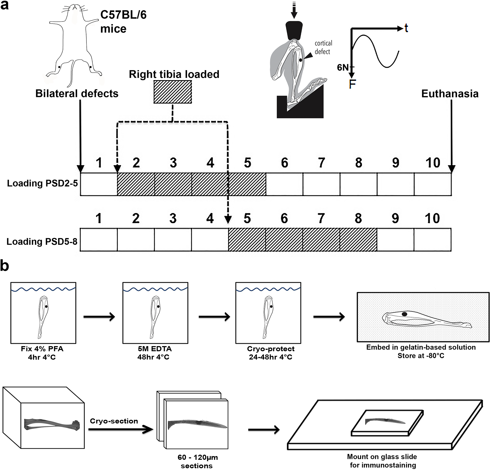
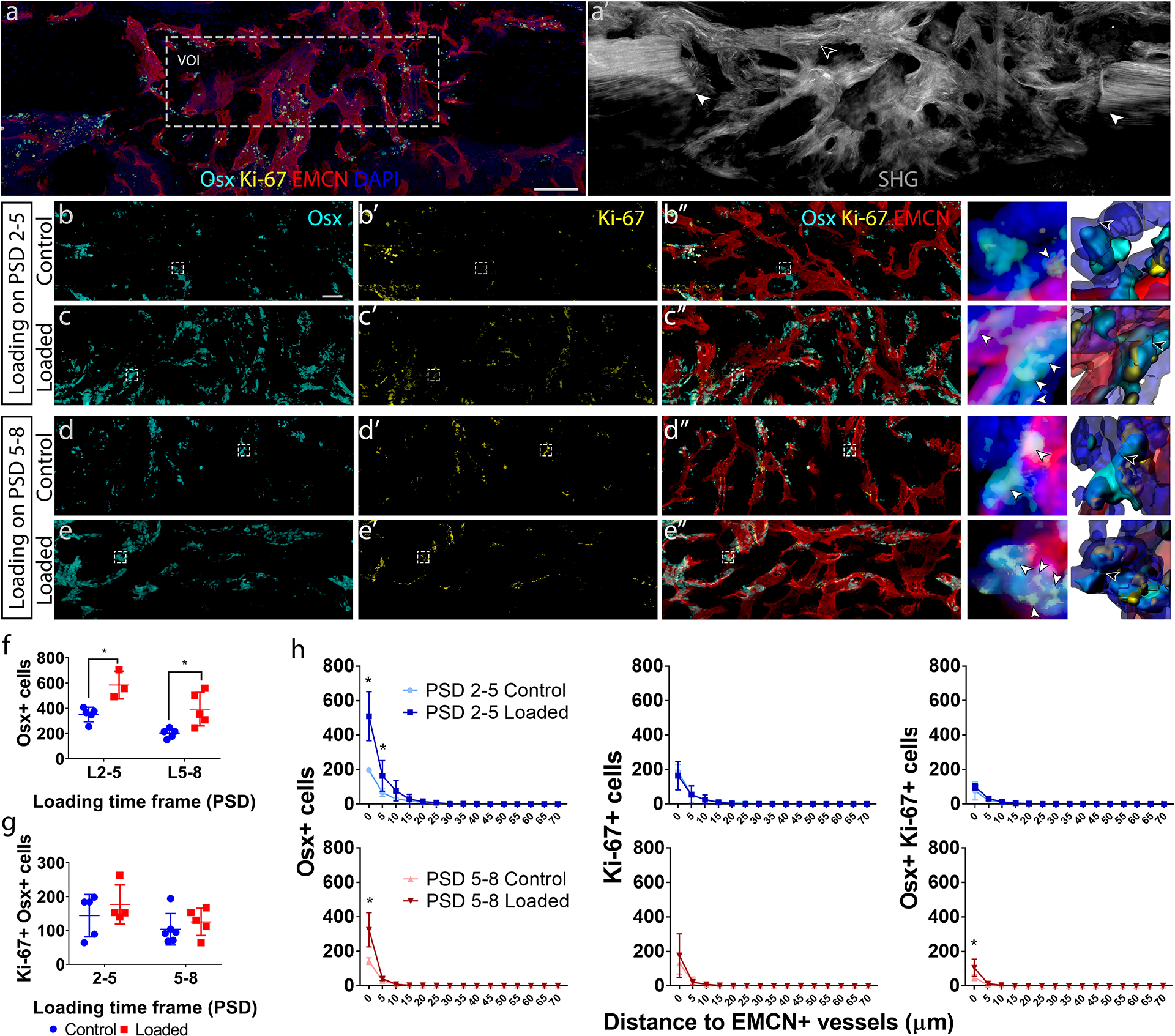
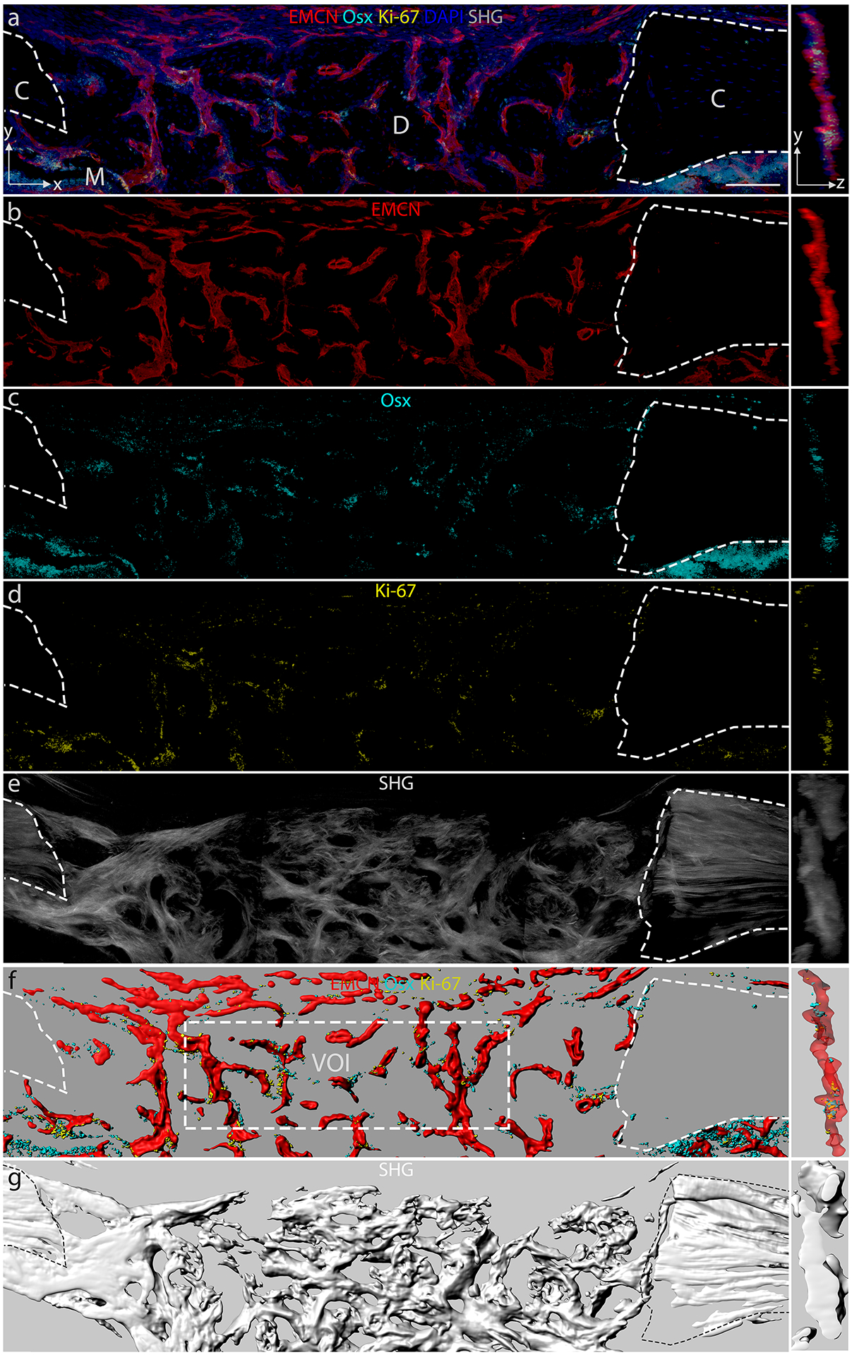
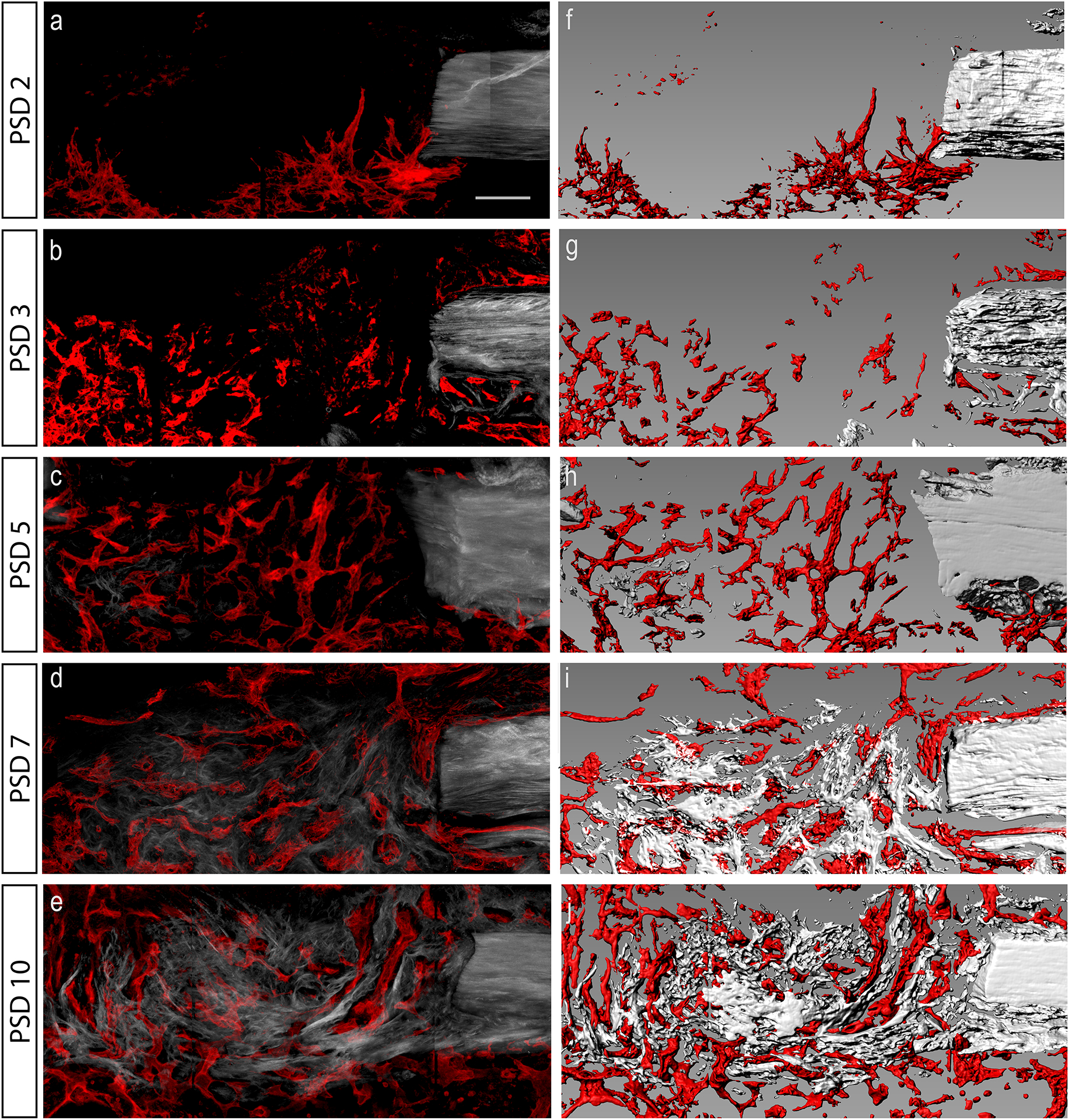
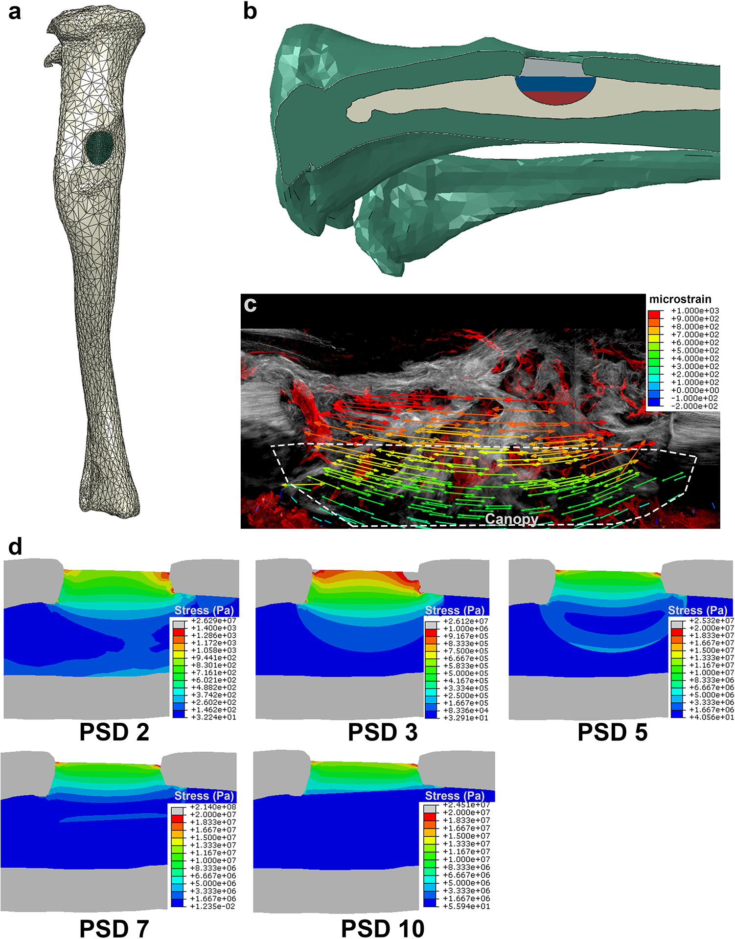
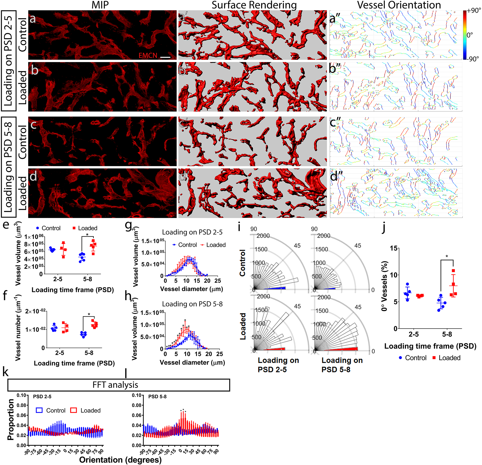
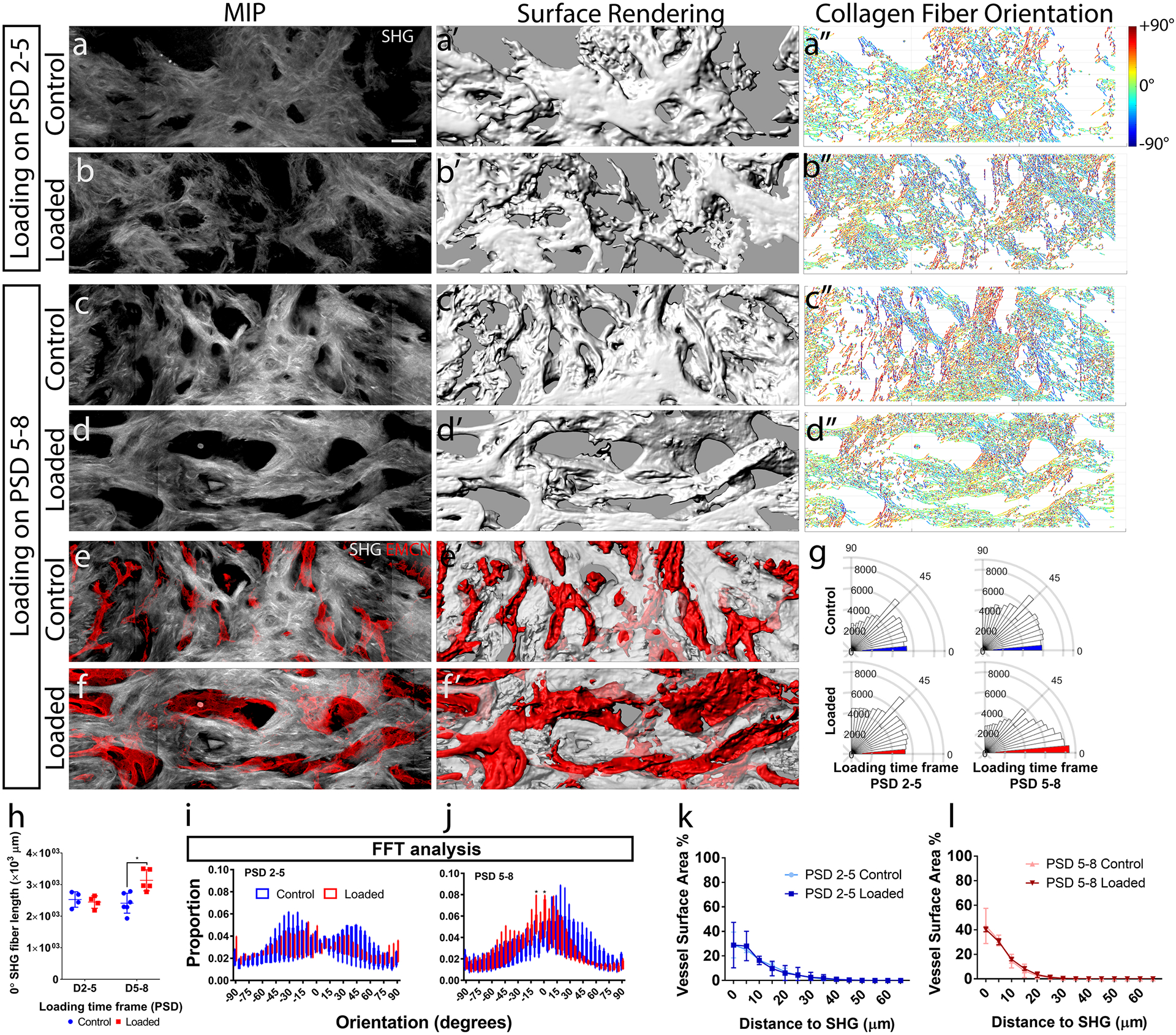
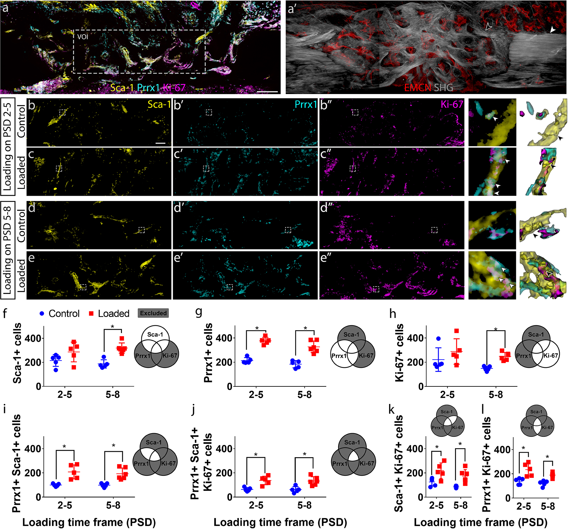
Elucidating the effects of mechanical stimulation on bone repair is crucial for optimization of the healing process. Specifically, the regulatory role that mechanical loading exerts on the osteogenic stem cell pool and vascular morphology during healing is incompletely understood. Because dynamic loading has been shown to enhance osteogenesis and repair, we hypothesized that loading induces the expansion of the osteoprogenitor cell population within a healing bone defect, leading to an increased presence of osteogenic cells. We further hypothesized that loading during the repair process regulates vascular and collagen matrix morphology and spatial interactions between vessels and osteogenic cells. To address these hypotheses, we used a mechanobiological bone repair model, which produces a consistent and reproducible intramembranous repair response confined in time and space. Bilateral tibial defects were created in adult C57BL/6 mice, which were subjected to axial compressive dynamic loading either during the early cellular invasion phase on postsurgical days (PSDs) 2 to 5 or during the matrix deposition phase on PSD 5 to 8. Confocal and two-photon microscopy was used to generate high-resolution three-dimensional (3D) renderings of longitudinal thick sections of the defect on PSD 10. Endomucin (EMCN)-positive vessels, Paired related homeobox 1 (Prrx1+) stem cell antigen-1 positive (Sca-1+) primitive osteoprogenitors (OPCs), and osterix positive (Osx+) preosteoblasts were visualized and quantified using deep tissue immunohistochemistry. New bone matrix was visualized with second harmonic generation autofluorescence of collagen fibers. We found that mechanical loading during the matrix deposition phase (PSD 5 to 8) increased vessel volume and number, and aligned vessels and collagen fibers to the load-bearing direction of bone. Furthermore, loading led to a significant increase in the proliferation and number of Prrx1+ Sca-1+ primitive OPCs, but not Osx+ preosteoblasts within the defect. Together, these data illustrate the adaptation of both collagen matrix and vascular morphology to better withstand mechanical load during bone repair, and that the mechanoresponsive cell population consists of the primitive osteogenic progenitors. © 2019 American Society for Bone and Mineral Research.
阐明机械刺激对骨修复的影响对于优化愈合过程至关重要。具体而言,机械负荷在愈合过程中对成骨干细胞池和血管形态的调节作用尚未完全明确。由于动态负荷已被证明可促进成骨和修复,我们推测负荷可诱导愈合骨缺损内骨祖细胞群体的扩增,从而导致成骨细胞数量增加。我们进一步推测,修复过程中的负荷可调节血管和胶原基质形态以及血管与成骨细胞之间的空间相互作用。为了验证这些假设,我们使用了一种机械生物学骨修复模型,该模型可在时间和空间上产生一致且可重复的膜内修复反应。在成年C57BL/6小鼠中制造双侧胫骨缺损,在术后第2至5天的早期细胞侵入阶段或术后第5至8天的基质沉积阶段对其施加轴向压缩动态负荷。使用共聚焦和双光子显微镜对术后第10天缺损的纵向厚切片进行高分辨率三维(3D)重建。使用深部组织免疫组织化学对内皮黏液素(EMCN)阳性血管、配对相关同源盒1(Prrx1+)干细胞抗原-1阳性(Sca-1+)原始骨祖细胞(OPC)和osterix阳性(Osx+)前成骨细胞进行可视化和定量分析。用胶原纤维的二次谐波产生自发荧光观察新骨基质。我们发现,在基质沉积阶段(术后第5至8天)进行机械负荷可增加血管体积和数量,并使血管和胶原纤维沿骨的承重方向排列。此外,负荷导致缺损内Prrx1+ Sca-1+原始OPC的增殖和数量显著增加,但Osx+前成骨细胞数量未增加。总之,这些数据表明,在骨修复过程中,胶原基质和血管形态均发生适应性变化,以更好地承受机械负荷,且机械反应性细胞群体由原始成骨祖细胞组成。©2019美国骨与矿物质研究学会。