UTS-SUSTech Joint Research Centre for Biomedical Materials and Devices, Department of Biomedical Engineering, Southern University of Science and Technology, Shenzhen, China.
Institute for Biomedical Materials and Devices (IBMD), Faculty of Science, University of Technology Sydney, Sydney, NSW, Australia.
Nat Commun. 2023 Oct 9;14(1):6287. doi: 10.1038/s41467-023-42001-2.
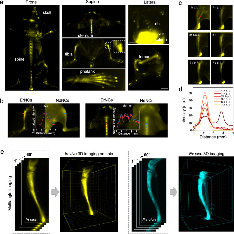
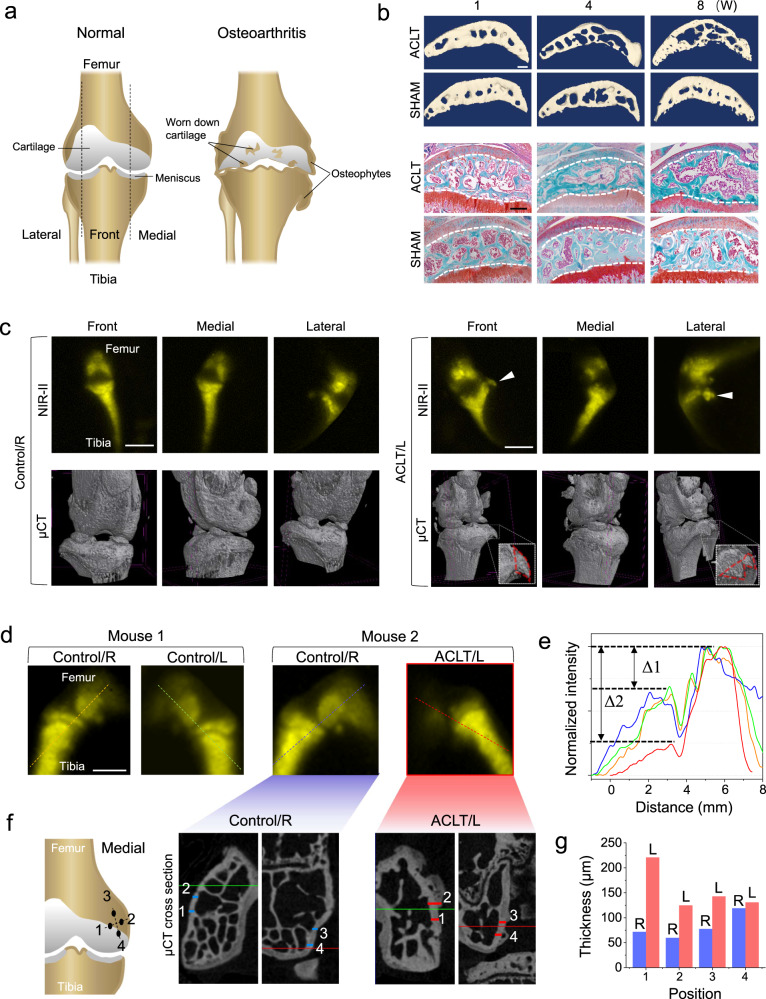
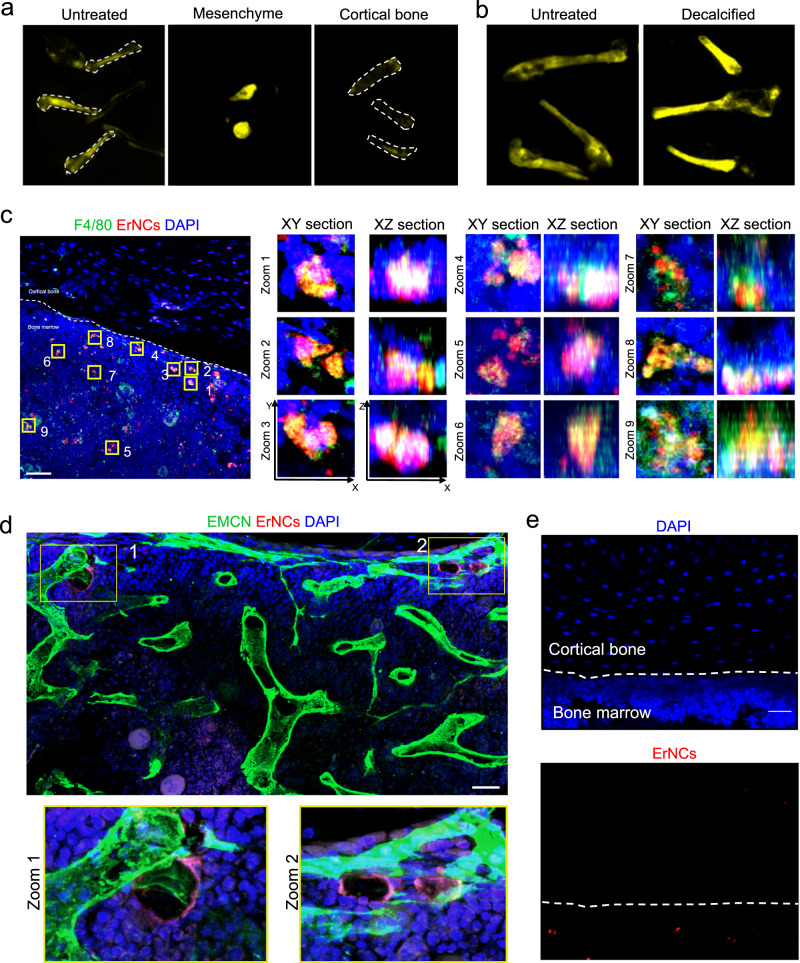
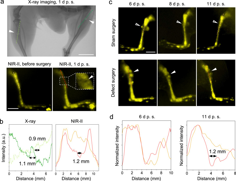
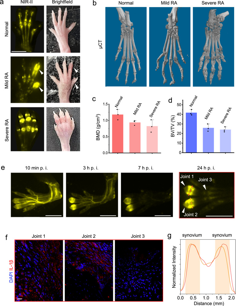
Skeletal disorders are commonly diagnosed by X-ray imaging, but the radiation limits its use. Optical imaging through the near-infrared-II window (NIR-II, 1000-1700 nm) can penetrate deep tissues without radiation risk, but the targeting of contrast agent is non-specific. Here, we report that lanthanide-doped nanocrystals can passively target the bone marrow, which can be effective for over two months. We therefore develop the high-resolution NIR-II imaging method for bone disease diagnosis, including the 3D bone imaging instrumentation to show the intravital bone morphology. We demonstrate the monitoring of 1 mm bone defects with spatial resolution comparable to the X-ray imaging result. Moreover, NIR-II imaging can reveal the early onset inflammation as the synovitis in the early stage of rheumatoid arthritis, comparable to micro computed tomography (μCT) in diagnosis of osteoarthritis, including the symptoms of osteophyte and hyperostosis in the knee joint.
骨骼疾病通常通过 X 射线成像来诊断,但辐射限制了其使用。近红外二区(NIR-II,1000-1700nm)的光学成像是无辐射风险的,可以穿透深部组织,但对比剂的靶向是非特异性的。在这里,我们报告镧系掺杂纳米晶体可以被动靶向骨髓,这对骨疾病的诊断具有长达两个月以上的有效作用。因此,我们开发了用于骨疾病诊断的高分辨率 NIR-II 成像方法,包括用于显示活体骨形态的 3D 骨成像仪器。我们展示了具有与 X 射线成像结果相当的空间分辨率的 1mm 骨缺损监测。此外,NIR-II 成像可以揭示早期炎症,如类风湿关节炎早期的滑膜炎,在诊断骨关节炎方面可与微计算机断层扫描(μCT)相媲美,包括膝关节的骨赘和骨质增生等症状。