• 文献检索
  • 文档翻译
  • 深度研究
  • 学术资讯
  • Suppr Zotero 插件Zotero 插件
  • 邀请有礼
  • 套餐&价格
  • 历史记录
应用&插件
Suppr Zotero 插件Zotero 插件浏览器插件Mac 客户端Windows 客户端微信小程序
定价
高级版会员购买积分包购买API积分包
服务
文献检索文档翻译深度研究API 文档MCP 服务
关于我们
关于 Suppr公司介绍联系我们用户协议隐私条款
关注我们

Suppr 超能文献

核心技术专利:CN118964589B侵权必究
粤ICP备2023148730 号-1Suppr @ 2026

文献检索

告别复杂PubMed语法,用中文像聊天一样搜索,搜遍4000万医学文献。AI智能推荐,让科研检索更轻松。

立即免费搜索

文件翻译

保留排版,准确专业,支持PDF/Word/PPT等文件格式,支持 12+语言互译。

免费翻译文档

深度研究

AI帮你快速写综述,25分钟生成高质量综述,智能提取关键信息,辅助科研写作。

立即免费体验

朊病毒病期间的神经炎症、小胶质细胞和细胞关联。

Neuroinflammation, Microglia, and Cell-Association during Prion Disease.

机构信息

Laboratory of Persistent Viral Diseases, Rocky Mountain Laboratories, National Institute of Allergy and Infectious Diseases, National Institutes of Health, Hamilton, MT 59840, USA.

出版信息

Viruses. 2019 Jan 15;11(1):65. doi: 10.3390/v11010065.

DOI:10.3390/v11010065
PMID:30650564
原文链接:https://pmc.ncbi.nlm.nih.gov/articles/PMC6356204/
Abstract

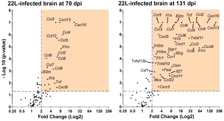
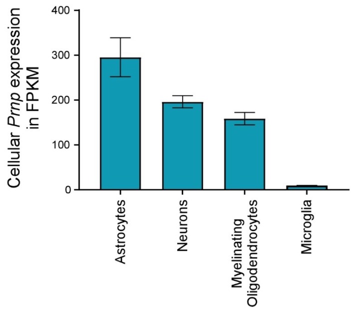
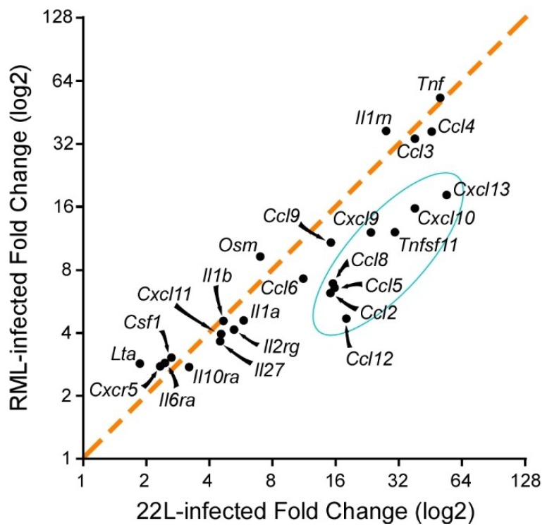
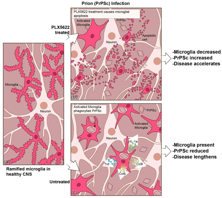
Prion disorders are transmissible diseases caused by a proteinaceous infectious agent that can infect the lymphatic and nervous systems. The clinical features of prion diseases can vary, but common hallmarks in the central nervous system (CNS) are deposition of abnormally folded protease-resistant prion protein (PrPres or PrPSc), astrogliosis, microgliosis, and neurodegeneration. Numerous proinflammatory effectors expressed by astrocytes and microglia are increased in the brain during prion infection, with many of them potentially damaging to neurons when chronically upregulated. Microglia are important first responders to foreign agents and damaged cells in the CNS, but these immune-like cells also serve many essential functions in the healthy CNS. Our current understanding is that microglia are beneficial during prion infection and critical to host defense against prion disease. Studies indicate that reduction of the microglial population accelerates disease and increases PrPSc burden in the CNS. Thus, microglia are unlikely to be a foci of prion propagation in the brain. In contrast, neurons and astrocytes are known to be involved in prion replication and spread. Moreover, certain astrocytes, such as A1 reactive astrocytes, have proven neurotoxic in other neurodegenerative diseases, and thus might also influence the progression of prion-associated neurodegeneration.

摘要

朊病毒疾病是由可感染淋巴和神经系统的蛋白质传染性病原体引起的传染性疾病。朊病毒疾病的临床特征可能有所不同,但中枢神经系统(CNS)的常见特征是异常折叠的蛋白酶抗性朊病毒蛋白(PrPres 或 PrPSc)、星形胶质细胞增生、小胶质细胞增生和神经退行性变的沉积。朊病毒感染期间,大脑中表达的许多促炎效应物增加,其中许多在慢性上调时对神经元具有潜在的破坏性。小胶质细胞是中枢神经系统中外来物和受损细胞的重要第一反应者,但这些免疫样细胞在健康的中枢神经系统中也具有许多重要功能。我们目前的理解是,小胶质细胞在朊病毒感染期间是有益的,并且对宿主防御朊病毒疾病至关重要。研究表明,减少小胶质细胞群体会加速疾病并增加 CNS 中的 PrPSc 负担。因此,小胶质细胞不太可能是大脑中朊病毒传播的焦点。相比之下,神经元和星形胶质细胞已知参与朊病毒的复制和传播。此外,某些星形胶质细胞,如 A1 反应性星形胶质细胞,已被证明在其他神经退行性疾病中具有神经毒性,因此也可能影响朊病毒相关神经退行性变的进展。

https://cdn.ncbi.nlm.nih.gov/pmc/blobs/4926/6356204/52f24d68a8f5/viruses-11-00065-g006.jpg
https://cdn.ncbi.nlm.nih.gov/pmc/blobs/4926/6356204/5d96c47c29cf/viruses-11-00065-g001.jpg
https://cdn.ncbi.nlm.nih.gov/pmc/blobs/4926/6356204/6538718cbe0d/viruses-11-00065-g002.jpg
https://cdn.ncbi.nlm.nih.gov/pmc/blobs/4926/6356204/1710fb49c2e4/viruses-11-00065-g003.jpg
https://cdn.ncbi.nlm.nih.gov/pmc/blobs/4926/6356204/0e789c6c507d/viruses-11-00065-g004.jpg
https://cdn.ncbi.nlm.nih.gov/pmc/blobs/4926/6356204/caaceb71e38d/viruses-11-00065-g005.jpg
https://cdn.ncbi.nlm.nih.gov/pmc/blobs/4926/6356204/52f24d68a8f5/viruses-11-00065-g006.jpg

相似文献

1
Neuroinflammation, Microglia, and Cell-Association during Prion Disease.朊病毒病期间的神经炎症、小胶质细胞和细胞关联。
Viruses. 2019 Jan 15;11(1):65. doi: 10.3390/v11010065.
2
Microglia Are Critical in Host Defense against Prion Disease.小胶质细胞在宿主抵抗朊病毒病中起关键作用。
J Virol. 2018 Jul 17;92(15). doi: 10.1128/JVI.00549-18. Print 2018 Aug 1.
3
Complement 3-astrocytes are highly abundant in prion diseases, but their abolishment led to an accelerated disease course and early dysregulation of microglia.补体 3-星形胶质细胞在朊病毒病中高度丰富,但它们的缺失导致疾病进程加速和小胶质细胞的早期失调。
Acta Neuropathol Commun. 2019 May 22;7(1):83. doi: 10.1186/s40478-019-0735-1.
4
RNA-seq and network analysis reveal unique glial gene expression signatures during prion infection.RNA-seq 和网络分析揭示朊病毒感染过程中独特的神经胶质基因表达特征。
Mol Brain. 2020 May 7;13(1):71. doi: 10.1186/s13041-020-00610-8.
5
Role of cyclophilin A from brains of prion-infected mice in stimulation of cytokine release by microglia and astroglia in vitro.朊病毒感染小鼠脑中亲环素 A 在体外刺激小胶质细胞和星形胶质细胞细胞因子释放中的作用。
J Biol Chem. 2012 Feb 10;287(7):4628-39. doi: 10.1074/jbc.M111.269480. Epub 2011 Dec 16.
6
Microglia in prion diseases.朊病毒疾病中的小胶质细胞。
J Clin Invest. 2017 Sep 1;127(9):3230-3239. doi: 10.1172/JCI90605. Epub 2017 Jul 17.
7
Flow Cytometric Detection of PrP in Neurons and Glial Cells from Prion-Infected Mouse Brains.朊病毒感染小鼠大脑中神经元和神经胶质细胞中朊蛋白的流式细胞术检测
J Virol. 2017 Dec 14;92(1). doi: 10.1128/JVI.01457-17. Print 2018 Jan 1.
8
Inflammatory response of microglia to prions is controlled by sialylation of PrP.小胶质细胞对朊病毒的炎症反应受 PrP 唾液酸化的控制。
Sci Rep. 2018 Jul 27;8(1):11326. doi: 10.1038/s41598-018-29720-z.
9
Activation of microglia by retroviral infection correlates with transient clearance of prions from the brain but does not change incubation time.逆转录病毒感染激活小胶质细胞与朊病毒从大脑中的短暂清除相关,但不改变潜伏期。
Brain Pathol. 2017 Sep;27(5):590-602. doi: 10.1111/bpa.12441. Epub 2016 Nov 21.
10
Prion protein expression differences in microglia and astroglia influence scrapie-induced neurodegeneration in the retina and brain of transgenic mice.小胶质细胞和星形胶质细胞中朊病毒蛋白表达差异影响转基因小鼠视网膜和大脑中的羊瘙痒病诱导的神经退行性变。
J Virol. 2007 Oct;81(19):10340-51. doi: 10.1128/JVI.00865-07. Epub 2007 Jul 25.

引用本文的文献

1
Knockout of P2Y12 receptor facilitates neuronal envelopment by reactive microglia and accelerates prion disease.P2Y12受体基因敲除促进反应性小胶质细胞对神经元的包裹并加速朊病毒病。
J Neuroinflammation. 2025 Aug 31;22(1):210. doi: 10.1186/s12974-025-03542-z.
2
Microglia-specific NF-κB signaling is a critical regulator of prion-induced glial inflammation and neuronal loss.小胶质细胞特异性核因子κB信号通路是朊病毒诱导的胶质细胞炎症和神经元损失的关键调节因子。
PLoS Pathog. 2025 Jun 18;21(6):e1012582. doi: 10.1371/journal.ppat.1012582. eCollection 2025 Jun.
3
Neuroinflammation-A Crucial Factor in the Pathophysiology of Depression-A Comprehensive Review.

本文引用的文献

1
Toll-like receptor 2 confers partial neuroprotection during prion disease.Toll 样受体 2 在朊病毒病中提供部分神经保护。
PLoS One. 2018 Dec 31;13(12):e0208559. doi: 10.1371/journal.pone.0208559. eCollection 2018.
2
Microglia Are Critical in Host Defense against Prion Disease.小胶质细胞在宿主抵抗朊病毒病中起关键作用。
J Virol. 2018 Jul 17;92(15). doi: 10.1128/JVI.00549-18. Print 2018 Aug 1.
3
Temporal Tracking of Microglia Activation in Neurodegeneration at Single-Cell Resolution.单细胞分辨率下神经退行性变中小胶质细胞激活的时间追踪。
神经炎症——抑郁症病理生理学中的关键因素——综述
Biomolecules. 2025 Mar 30;15(4):502. doi: 10.3390/biom15040502.
4
Histone deacetylases: the critical enzymes for microglial activation involved in neuropathic pain.组蛋白去乙酰化酶:参与神经性疼痛的小胶质细胞激活的关键酶
Front Pharmacol. 2025 Mar 6;16:1515787. doi: 10.3389/fphar.2025.1515787. eCollection 2025.
5
Therapeutic effect of curcumin derivative GT863 on prion-infected mice.姜黄素衍生物GT863对朊病毒感染小鼠的治疗作用。
Sci Rep. 2025 Feb 14;15(1):5444. doi: 10.1038/s41598-025-89317-1.
6
Limbic system synaptic dysfunctions associated with prion disease onset.与朊病毒病发病相关的边缘系统突触功能障碍。
Acta Neuropathol Commun. 2024 Dec 20;12(1):192. doi: 10.1186/s40478-024-01905-w.
7
Diagnostic and Prognostic Value of Plasma GFAP in Sporadic Creutzfeldt-Jakob Disease in the Clinical Setting of Rapidly Progressive Dementia.在快速进展性痴呆的临床环境中,血浆 GFAP 在散发性克雅氏病中的诊断和预后价值。
Int J Mol Sci. 2024 May 8;25(10):5106. doi: 10.3390/ijms25105106.
8
Interactions between Cytokines and the Pathogenesis of Prion Diseases: Insights and Implications.细胞因子与朊病毒疾病发病机制之间的相互作用:见解与启示
Brain Sci. 2024 Apr 23;14(5):413. doi: 10.3390/brainsci14050413.
9
The Role of Glial Cells in Neurobiology and Prion Neuropathology.胶质细胞在神经生物学和朊病毒神经病理学中的作用。
Cells. 2024 May 14;13(10):832. doi: 10.3390/cells13100832.
10
CD11c is not required by microglia to convey neuroprotection after prion infection.CD11c 并非小胶质细胞在朊病毒感染后发挥神经保护作用所必需的。
PLoS One. 2023 Nov 1;18(11):e0293301. doi: 10.1371/journal.pone.0293301. eCollection 2023.
Cell Rep. 2017 Oct 10;21(2):366-380. doi: 10.1016/j.celrep.2017.09.039.
4
Microglia emerge as central players in brain disease.小胶质细胞成为大脑疾病的核心参与者。
Nat Med. 2017 Sep 8;23(9):1018-1027. doi: 10.1038/nm.4397.
5
Statins are ineffective at reducing neuroinflammation or prolonging survival in scrapie-infected mice.他汀类药物在降低瘙痒病感染小鼠的神经炎症或延长其生存期方面无效。
J Gen Virol. 2017 Aug;98(8):2190-2199. doi: 10.1099/jgv.0.000876. Epub 2017 Jul 31.
6
Microglia in prion diseases.朊病毒疾病中的小胶质细胞。
J Clin Invest. 2017 Sep 1;127(9):3230-3239. doi: 10.1172/JCI90605. Epub 2017 Jul 17.
7
Reactive Astrocytes: Production, Function, and Therapeutic Potential.反应性星形胶质细胞:产生、功能和治疗潜力。
Immunity. 2017 Jun 20;46(6):957-967. doi: 10.1016/j.immuni.2017.06.006.
8
α-synuclein toxicity in neurodegeneration: mechanism and therapeutic strategies.神经退行性变中的α-突触核蛋白毒性:机制与治疗策略
Nat Med. 2017 Feb 7;23(2):1-13. doi: 10.1038/nm.4269.
9
Neurotoxic reactive astrocytes are induced by activated microglia.神经毒性反应性星形胶质细胞由活化的小胶质细胞诱导产生。
Nature. 2017 Jan 26;541(7638):481-487. doi: 10.1038/nature21029. Epub 2017 Jan 18.
10
How neuroinflammation contributes to neurodegeneration.神经炎症如何导致神经退行性变。
Science. 2016 Aug 19;353(6301):777-83. doi: 10.1126/science.aag2590.