Laboratory of Clinical and Experimental Endocrinology, Department of Chronic Diseases, Metabolism and Ageing, KU Leuven, Leuven, Belgium.
Laboratory of Angiogenesis and Vascular Biology, VIB Center for Cancer Biology, VIB, Leuven, Belgium.
Nature. 2019 Jan;565(7740):511-515. doi: 10.1038/s41586-019-0874-3. Epub 2019 Jan 16.
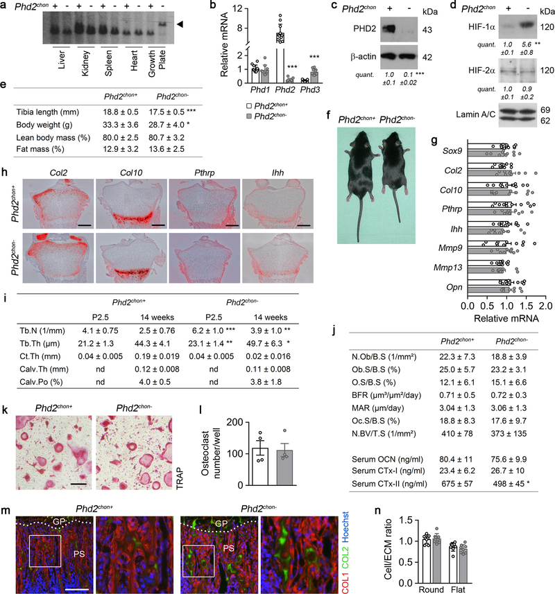
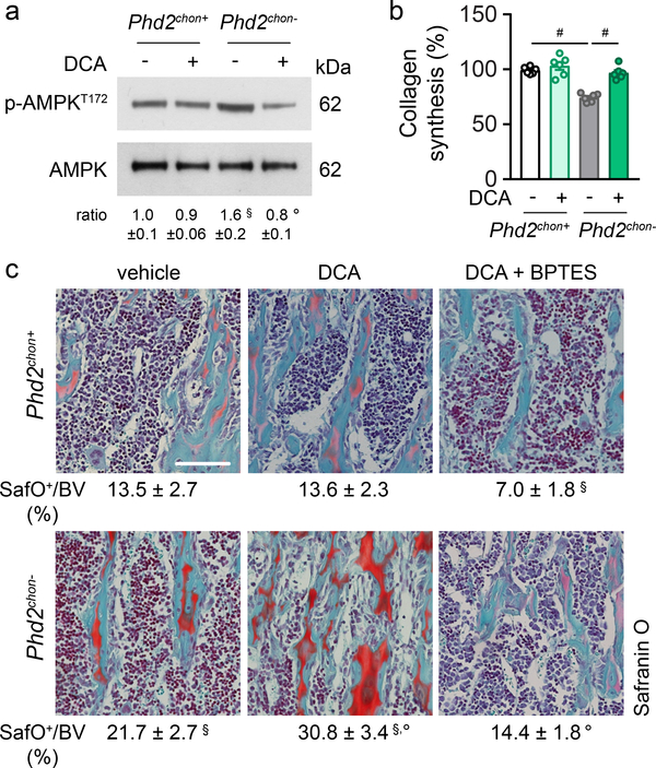
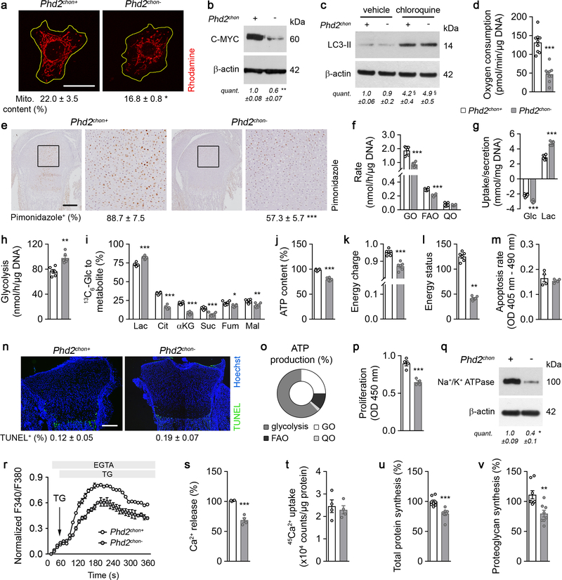
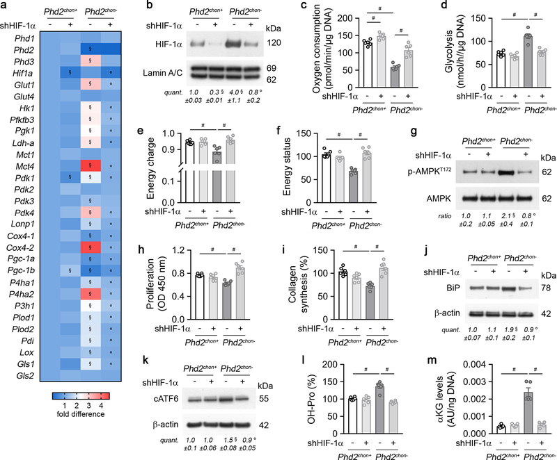
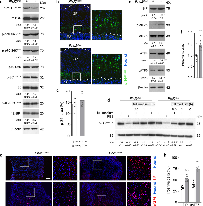
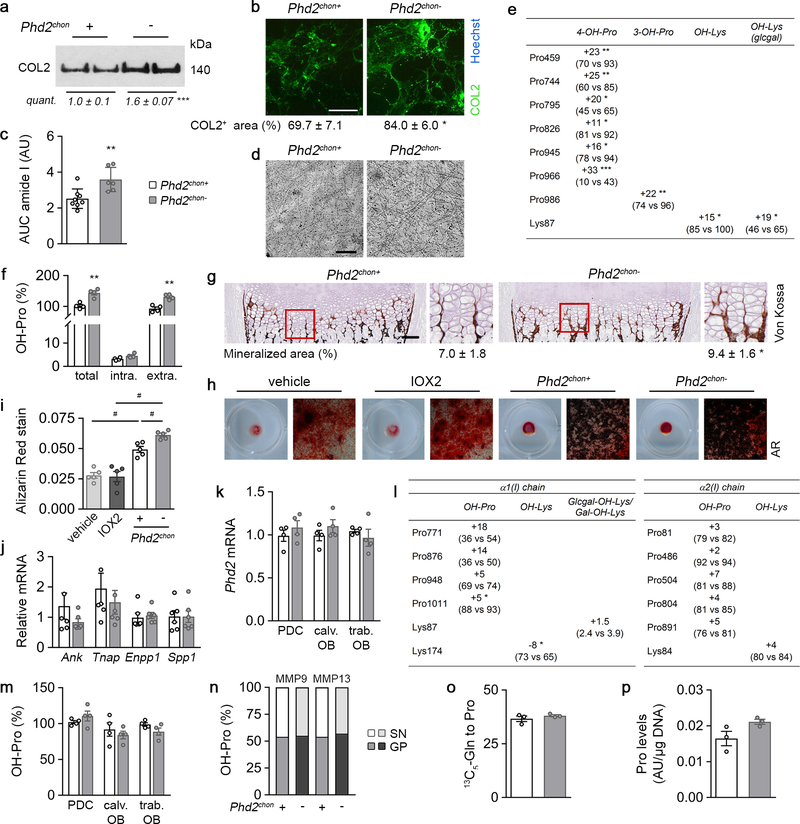
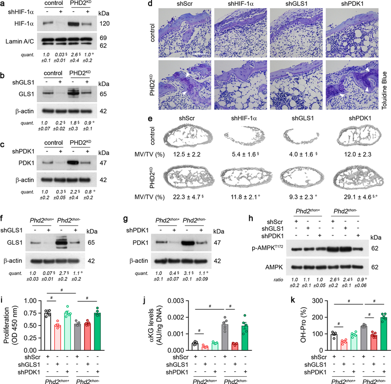
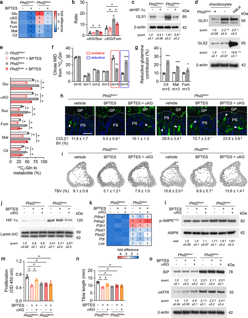
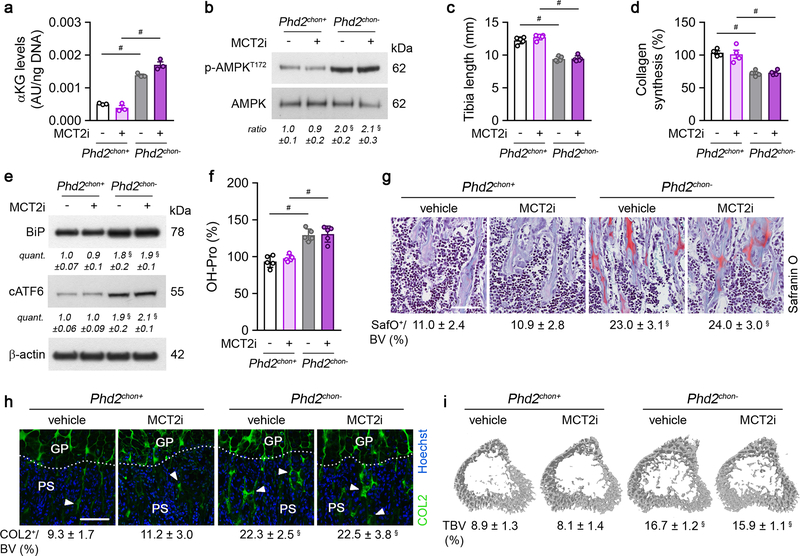
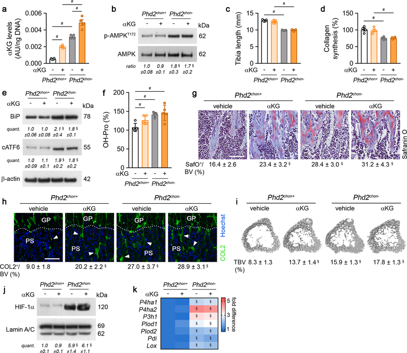
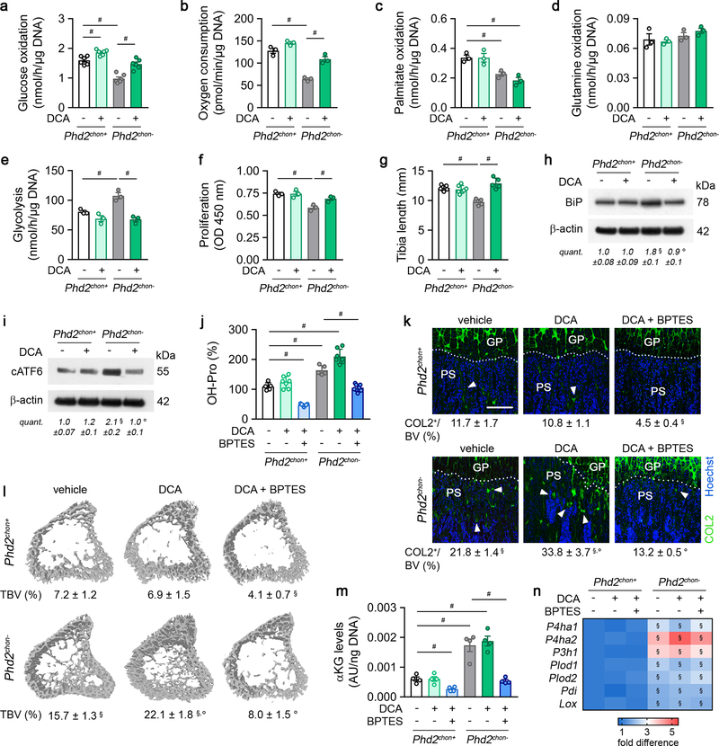
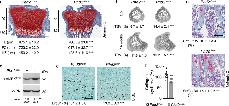
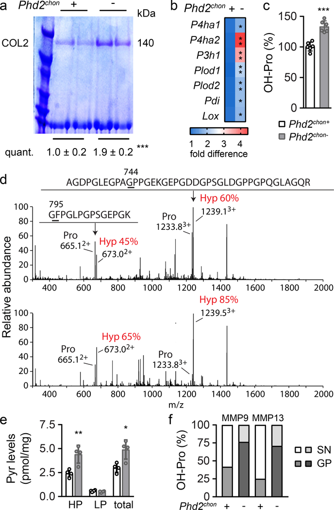
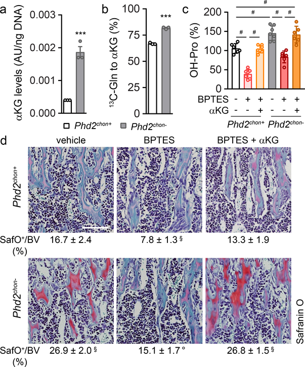
Endochondral ossification, an important process in vertebrate bone formation, is highly dependent on correct functioning of growth plate chondrocytes. Proliferation of these cells determines longitudinal bone growth and the matrix deposited provides a scaffold for future bone formation. However, these two energy-dependent anabolic processes occur in an avascular environment. In addition, the centre of the expanding growth plate becomes hypoxic, and local activation of the hypoxia-inducible transcription factor HIF-1α is necessary for chondrocyte survival by unidentified cell-intrinsic mechanisms. It is unknown whether there is a requirement for restriction of HIF-1α signalling in the other regions of the growth plate and whether chondrocyte metabolism controls cell function. Here we show that prolonged HIF-1α signalling in chondrocytes leads to skeletal dysplasia by interfering with cellular bioenergetics and biosynthesis. Decreased glucose oxidation results in an energy deficit, which limits proliferation, activates the unfolded protein response and reduces collagen synthesis. However, enhanced glutamine flux increases α-ketoglutarate levels, which in turn increases proline and lysine hydroxylation on collagen. This metabolically regulated collagen modification renders the cartilaginous matrix more resistant to protease-mediated degradation and thereby increases bone mass. Thus, inappropriate HIF-1α signalling results in skeletal dysplasia caused by collagen overmodification, an effect that may also contribute to other diseases involving the extracellular matrix such as cancer and fibrosis.
软骨内骨化是脊椎动物骨骼形成的一个重要过程,高度依赖于生长板软骨细胞的正常功能。这些细胞的增殖决定了骨骼的纵向生长,而沉积的基质为未来的骨骼形成提供了支架。然而,这两个依赖能量的合成代谢过程发生在无血管的环境中。此外,不断扩大的生长板中心变得缺氧,局部激活缺氧诱导转录因子 HIF-1α 对于软骨细胞的存活是必要的,但其具体机制尚不清楚。目前尚不清楚在生长板的其他区域是否需要限制 HIF-1α 信号,以及软骨细胞代谢是否控制细胞功能。在这里,我们表明,软骨细胞中 HIF-1α 信号的持续激活会通过干扰细胞的生物能量和生物合成导致骨骼发育不良。葡萄糖氧化减少会导致能量不足,从而限制增殖,激活未折叠蛋白反应并减少胶原蛋白合成。然而,增强的谷氨酰胺通量会增加α-酮戊二酸水平,进而增加胶原蛋白上脯氨酸和赖氨酸的羟化。这种代谢调节的胶原蛋白修饰使软骨基质更能抵抗蛋白酶介导的降解,从而增加骨量。因此,不适当的 HIF-1α 信号会导致胶原蛋白过度修饰引起的骨骼发育不良,这种效应可能也与涉及细胞外基质的其他疾病有关,如癌症和纤维化。