Mohr Corinna J, Steudel Friederike A, Gross Dominic, Ruth Peter, Lo Wing-Yee, Hoppe Reiner, Schroth Werner, Brauch Hiltrud, Huber Stephan M, Lukowski Robert
Department of Pharmacology, Toxicology and Clinical Pharmacy, Institute of Pharmacy, University of Tuebingen, 72076 Tuebingen, Germany.
Dr. Margarete Fischer-Bosch-Institute of Clinical Pharmacology, 70376 Stuttgart and University of Tuebingen, 72076 Tuebingen, Germany.
Cancers (Basel). 2019 Jan 17;11(1):109. doi: 10.3390/cancers11010109.
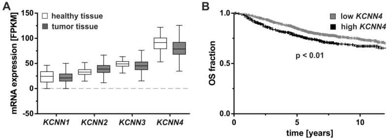
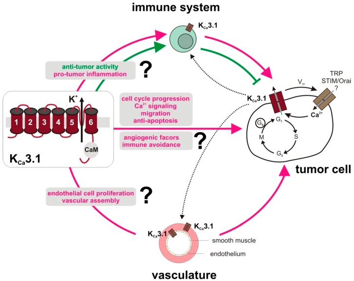
Several tumor entities have been reported to overexpress K3.1 potassium channels due to epigenetic, transcriptional, or post-translational modifications. By modulating membrane potential, cell volume, or Ca signaling, K3.1 has been proposed to exert pivotal oncogenic functions in tumorigenesis, malignant progression, metastasis, and therapy resistance. Moreover, K3.1 is expressed by tumor-promoting stroma cells such as fibroblasts and the tumor vasculature suggesting a role of K3.1 in the adaptation of the tumor microenvironment. Combined, this features K3.1 as a candidate target for innovative anti-cancer therapy. However, immune cells also express K3.1 thereby contributing to T cell activation. Thus, any strategy targeting K3.1 in anti-cancer therapy may also modulate anti-tumor immune activity and/or immunosuppression. The present review article highlights the potential of K3.1 as an anti-tumor target providing an overview of the current knowledge on its function in tumor pathogenesis with emphasis on vasculo- and angiogenesis as well as anti-cancer immune responses.
据报道,由于表观遗传、转录或翻译后修饰,几种肿瘤实体过表达K3.1钾通道。通过调节膜电位、细胞体积或钙信号,K3.1被认为在肿瘤发生、恶性进展、转移和治疗抗性中发挥关键的致癌功能。此外,K3.1由肿瘤促进基质细胞如成纤维细胞和肿瘤脉管系统表达,提示K3.1在肿瘤微环境适应中的作用。综合来看,这使K3.1成为创新抗癌治疗的候选靶点。然而,免疫细胞也表达K3.1,从而促进T细胞活化。因此,任何在抗癌治疗中靶向K3.1的策略也可能调节抗肿瘤免疫活性和/或免疫抑制。本综述文章强调了K3.1作为抗肿瘤靶点的潜力,概述了目前关于其在肿瘤发病机制中的功能的知识,重点是血管生成和血管新生以及抗癌免疫反应。