Zhang Hui, Ma Yunxia, Wang Haibao, Xu Liyan, Yu Yongqiang
Department of Radiology, The First Affiliated Hospital of Anhui Medical University, Hefei, Anhui 230022, P.R. China.
Department of Otorhinolaryngology Head and Neck Surgery, The First Affiliated Hospital of Anhui Medical University, Hefei, Anhui 230022, P.R. China.
Oncol Lett. 2019 Feb;17(2):1826-1832. doi: 10.3892/ol.2018.9806. Epub 2018 Dec 6.
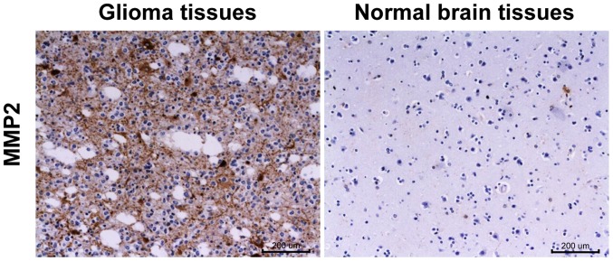
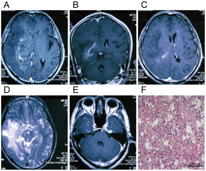
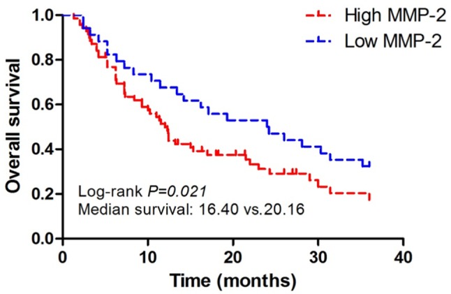
The expression of matrix metalloproteinase-2 (MMP-2) in brain glioma and its correlation with patients' clinicopathological characteristics and magnetic resonance imaging (MRI) features were investigated. A total of 104 patients with brain glioma admitted and treated in the First Affiliated Hospital of Anhui Medical University from June 2010 to September 2014 were randomly enrolled. MRI examination was performed before operation. Immunohistochemistry (IHC) was used to detect the expression levels of MMP-2 in brain glioma tissues and paired normal brain tissues after operation and to analyze the associations of MMP-2 expression with the clinicopathological characteristics of brain glioma and survival time of patients. The relationship between MMP-2 expression and preoperative MRI features of glioma was analyzed. The positive rate of MMP-2 expression in brain glioma was 73.08% (76/104), while that in paired normal brain tissues was only 12.5% (13/104), obviously lower than that in brain glioma tissues (P<0.05). The MMP-2 expression in the body of glioma was not related to the patients' sex, age, tumor location and pathological type (P>0.05), but there was a significant correlation with the tumor diameter and pathological grade of the patients (P<0.05). Analysis by Cox model suggested that tumor diameter, pathological grade and MMP-2 were independent prognostic factors for glioma (P<0.05). The overall survival (OS) of patients in the positive MMP-2 expression group was 16.4 months, while the OS in the negative MMP-2 expression group was 20.16 months, and the difference between the two groups was statistically significant (P<0.05). The positive expression of MMP-2 in glioma was closely related to the uniformity of MRI signal for tumor, tumor diameter, severity of peritumoral edema, degree of enhancement and pathological grade of tumor (P<0.05). MMP-2 is highly expressed in brain glioma, and it is a negative factor for prognosis. Therefore, the MRI manifestations of glioma can reflect to some extent the intensity of MMP-2 expression.
研究了基质金属蛋白酶-2(MMP-2)在脑胶质瘤中的表达及其与患者临床病理特征和磁共振成像(MRI)特征的相关性。随机纳入2010年6月至2014年9月在安徽医科大学第一附属医院收治的104例脑胶质瘤患者。术前进行MRI检查。采用免疫组织化学(IHC)检测术后脑胶质瘤组织及配对正常脑组织中MMP-2的表达水平,并分析MMP-2表达与脑胶质瘤临床病理特征及患者生存时间的相关性。分析MMP-2表达与胶质瘤术前MRI特征的关系。脑胶质瘤中MMP-2表达的阳性率为73.08%(76/104),而配对正常脑组织中的阳性率仅为12.5%(13/104),明显低于脑胶质瘤组织中的阳性率(P<0.05)。胶质瘤主体中MMP-2的表达与患者的性别、年龄、肿瘤位置及病理类型无关(P>0.05),但与患者的肿瘤直径及病理分级显著相关(P<0.05)。Cox模型分析表明,肿瘤直径、病理分级和MMP-2是胶质瘤的独立预后因素(P<0.05)。MMP-2表达阳性组患者的总生存期(OS)为16.4个月,而MMP-2表达阴性组的OS为20.16个月,两组间差异有统计学意义(P<0.05)。胶质瘤中MMP-2的阳性表达与肿瘤MRI信号均匀性、肿瘤直径、瘤周水肿严重程度、强化程度及肿瘤病理分级密切相关(P<0.05)。MMP-2在脑胶质瘤中高表达,是预后的负性因素。因此,胶质瘤的MRI表现可在一定程度上反映MMP-2表达的强度。