Institut Curie, PSL Research University, CNRS, UMR144, 26 rue d'Ulm, F-75005, Paris, France.
Sorbonne Universités, UPMC Paris 06, F-75005, Paris, France.
Breast Cancer Res. 2019 Jan 25;21(1):13. doi: 10.1186/s13058-019-1101-8.
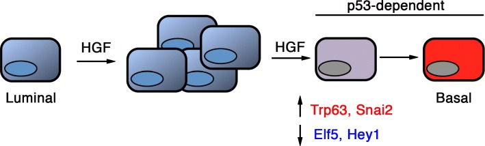
The adult mammary epithelium is composed of basal and luminal cells. The luminal lineage comprises two major cell populations, positive and negative for estrogen and progesterone receptors (ER and PR, respectively), both containing clonogenic progenitor cells. Deregulated ER/PR luminal progenitor cells are suspected to be at the origin of basal-type triple-negative (TNBC) breast cancers, a subtype frequently associated with loss of P53 function and MET signaling hyperactivation. Using mouse models, we recently reported that p53 restricts luminal progenitor cell amplification whereas paracrine Met activation stimulates their growth and favors a luminal-to-basal switch. Here, we analyzed how these two critical pathways interact to control luminal progenitor function.
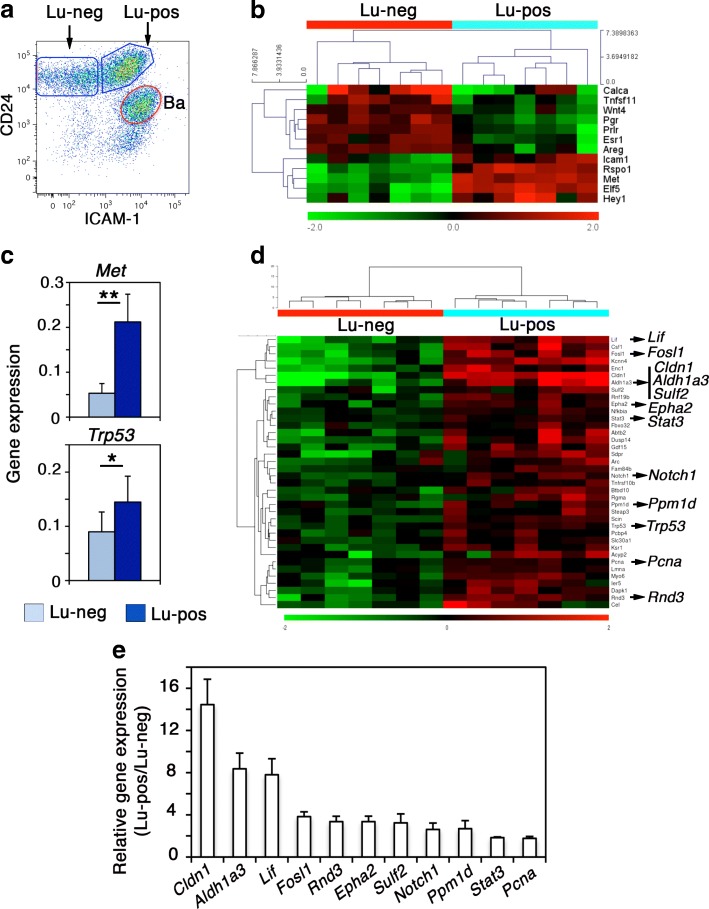
We have (i) established and analyzed the gene expression profile of luminal progenitors isolated by ICAM-1, a robust surface marker we previously identified; (ii) purified luminal progenitors from p53-deficient and p53-proficient mouse mammary epithelium to compare their functional and molecular characteristics; and (iii) analyzed their response to HGF, the major Met ligand, in three-dimensional cultures.
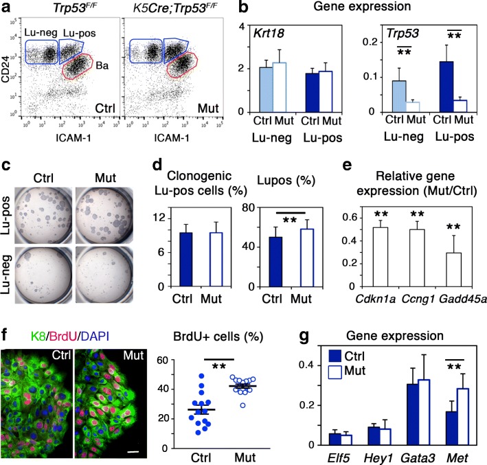
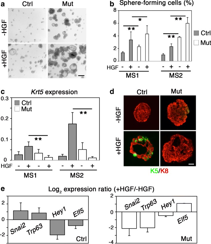
We found that luminal progenitors, compared to non-clonogenic luminal cells, overexpress Trp53 and numerous p53 target genes. In vivo, loss of Trp53 induced the expansion of luminal progenitors, affecting expression of several important p53 target genes including those encoding negative regulators of cell cycle progression. Consistently, p53-deficient luminal progenitors displayed increased proliferative and self-renewal activities in culture. However, they did not exhibit perturbed expression of luminal-specific markers and major regulators, such as Hey1, Elf5, and Gata3. Moreover, although expressing Met at higher level than p53-proficient luminal progenitors, p53-deficient luminal progenitors failed to acquire basal-specific features when stimulated by HGF, showing that p53 promotes the plastic behavior of luminal progenitors downstream of Met activation.
Our study reveals a crosstalk between Met- and p53-mediated signaling pathways in the regulation of luminal progenitor function. In particular, it shows that neither p53 loss alone nor p53 loss combined with Met signaling activation caused an early detectable cell fate alteration in luminal progenitors. Conceivably, additional events are required to confer basal-specific characteristics to luminal-derived TNBCs.
成人乳腺上皮由基底细胞和腔细胞组成。腔系由两种主要的细胞群体组成,分别对雌激素和孕激素受体(ER 和 PR,分别)呈阳性和阴性,两者都含有克隆形成祖细胞。失调的 ER/PR 腔前体细胞被怀疑是基底型三阴性(TNBC)乳腺癌的起源,这种亚型常与 p53 功能丧失和 MET 信号过度激活有关。我们最近使用小鼠模型报告称,p53 限制了腔前体细胞的扩增,而旁分泌 Met 激活刺激了它们的生长,并有利于从腔到基底的转变。在这里,我们分析了这两个关键途径如何相互作用以控制腔前体细胞的功能。
我们(i)通过我们之前鉴定的强大表面标志物 ICAM-1 分离和分析了腔前体细胞的基因表达谱;(ii)从 p53 缺陷和 p53 功能正常的小鼠乳腺上皮中纯化了腔前体细胞,以比较它们的功能和分子特征;(iii)分析了它们对 HGF(MET 的主要配体)在三维培养中的反应。
我们发现,与非克隆形成的腔细胞相比,腔前体细胞过度表达 Trp53 和许多 p53 靶基因。在体内,Trp53 的缺失诱导了腔前体细胞的扩增,影响了几个重要的 p53 靶基因的表达,包括那些编码细胞周期进程负调节剂的基因。一致地,p53 缺陷型腔前体细胞在培养中表现出增加的增殖和自我更新活性。然而,它们并没有表现出腔特异性标记物和主要调节因子(如 Hey1、Elf5 和 Gata3)表达的失调。此外,尽管 p53 缺陷型腔前体细胞表达的 Met 水平高于 p53 功能正常的腔前体细胞,但当受到 HGF 刺激时,它们未能获得基底特异性特征,表明 p53 促进了 Met 激活下游腔前体细胞的可塑性行为。
我们的研究揭示了 Met 和 p53 介导的信号通路在调节腔前体细胞功能中的相互作用。特别是,它表明 p53 缺失本身或 p53 缺失与 Met 信号激活相结合,在腔前体细胞中不会导致早期可检测的细胞命运改变。可以想象,赋予腔衍生的 TNBC 基底特异性特征需要额外的事件。