Creed Rose B, Goldberg Matthew S
Center for Neurodegeneration and Experimental Therapeutics, Department of Neurology, The University of Alabama at Birmingham, Birmingham, AL, United States.
Department of Neurobiology, The University of Alabama at Birmingham, Birmingham, AL, United States.
Front Neurosci. 2019 Jan 9;12:1034. doi: 10.3389/fnins.2018.01034. eCollection 2018.
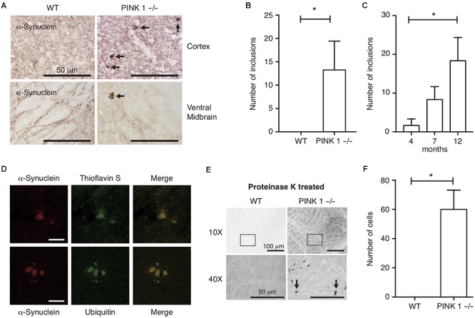
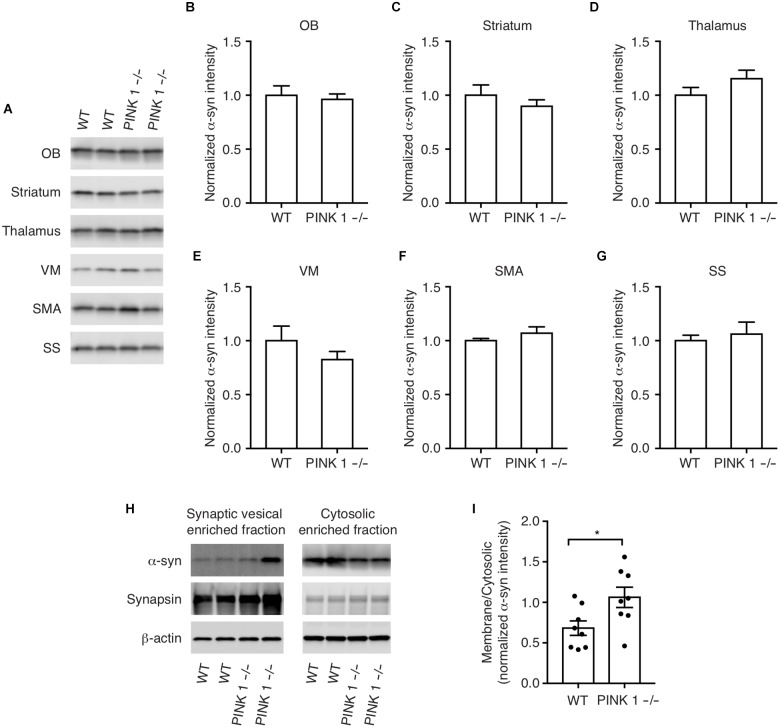
Mutations in PTEN induced kinase 1 (PINK1) cause autosomal recessive Parkinson's disease (PD). The main pathological hallmarks of PD are loss of dopaminergic neurons in the substantia nigra pars compacta and the formation of protein aggregates containing α-synuclein. Previous studies of PINK1 knockout (PINK1-/-) rats have reported mitochondrial dysfunction, locomotor behavioral deficits, loss of neurons in the substantia nigra and α-synuclein aggregates in various brain regions. We sought to characterize PINK1-/- rats in more detail specifically with respect to α-synuclein pathology because abnormal α-synuclein has been implicated genetically, biophysically and neuropathologically as a mechanism of PD pathogenesis. Moreover, the spontaneous formation of α-synuclein aggregates without α-synuclein overexpression, injection or toxin administration is a rare and important characteristic for an animal model of PD or other synucleinopathies, such as dementia with Lewy bodies and multiple system atrophy. We observed α-synuclein-immunoreactive aggregates in various brain regions of PINK1-/- rats including cortex, thalamus, striatum and ventral midbrain, but nowhere in wild-type (WT) rats. Co-immunofluorescence showed that the α-synuclein-immunoreactive aggregates are both thioflavin S and ubiquitin positive. Many cells in the brains of PINK1-/- rats but not WT rats contained protease-resistant α-synuclein. Total synuclein protein levels were unchanged; however, biochemical fractionation showed a significant shift of α-synuclein from the cytosolic fraction to the synaptic vesicle-enriched fraction of PINK1-/- brain homogenates compared to WT. This data indicates that PINK1 deficiency results in abnormal α-synuclein localization, protease resistance and aggregation . The PINK1-/- rat could be a useful animal model to study the role of abnormal α-synuclein in PD-related neurodegeneration.
磷酸酶与张力蛋白同源物缺失性诱导激酶1(PINK1)突变会导致常染色体隐性帕金森病(PD)。PD的主要病理特征是黑质致密部多巴胺能神经元缺失以及含有α-突触核蛋白的蛋白聚集体形成。先前对PINK1基因敲除(PINK1-/-)大鼠的研究报告了其线粒体功能障碍、运动行为缺陷、黑质神经元缺失以及不同脑区的α-突触核蛋白聚集体。我们试图更详细地描述PINK1-/-大鼠,特别是关于α-突触核蛋白病理学方面,因为异常的α-突触核蛋白在遗传、生物物理和神经病理学上被认为是PD发病机制之一。此外,在没有α-突触核蛋白过表达、注射或毒素给药的情况下自发形成α-突触核蛋白聚集体,对于PD或其他突触核蛋白病(如路易体痴呆和多系统萎缩)的动物模型来说是一种罕见且重要的特征。我们在PINK1-/-大鼠的不同脑区(包括皮质、丘脑、纹状体和腹侧中脑)观察到了α-突触核蛋白免疫反应性聚集体,但在野生型(WT)大鼠中未观察到。共免疫荧光显示,α-突触核蛋白免疫反应性聚集体对硫黄素S和泛素均呈阳性。PINK1-/-大鼠而非WT大鼠大脑中的许多细胞含有蛋白酶抗性α-突触核蛋白。总突触核蛋白水平未发生变化;然而,生化分级分离显示,与WT相比,PINK1-/-脑匀浆中α-突触核蛋白从胞质部分显著转移至富含突触小泡的部分。该数据表明PINK1缺乏会导致α-突触核蛋白定位异常、蛋白酶抗性和聚集。PINK1-/-大鼠可能是研究异常α-突触核蛋白在PD相关神经退行性变中作用的有用动物模型。