Center for Neurodegeneration and Experimental Therapeutics, The University of Alabama at Birmingham, Birmingham, AL, 35294, USA.
Department of Neurology, The University of Alabama at Birmingham, Birmingham, AL, 35294, USA.
Acta Neuropathol Commun. 2022 May 23;10(1):78. doi: 10.1186/s40478-022-01374-z.
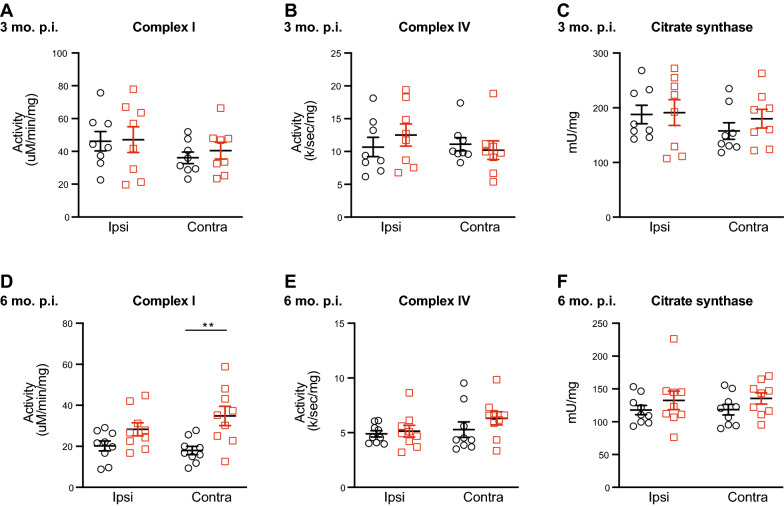
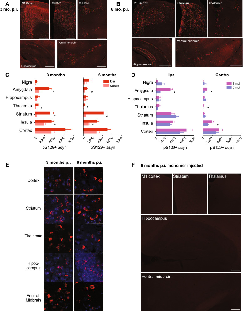
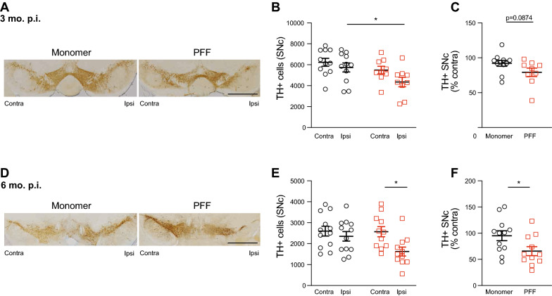
Genetic and neuropathological evidence strongly implicates aberrant forms of α-synuclein in neurodegeneration. Antibodies specific for α-synuclein phosphorylated at serine 129 (pS129) are selective for the pathological protein aggregates that are characteristic of Parkinson's disease (PD) and other synucleinopathies, such as dementia with Lewy bodies (DLB) and multiple system atrophy (MSA). Although the etiology of most synucleinopathies remains uncertain, a large body of evidence points to mitochondrial dysfunction. The recent development of animal models based on intracranial injection of α-synuclein pre-formed fibrils (PFFs) has provided a valuable experimental system in which to study the spread and neurotoxicity of α-synuclein aggregates, yet the effects of PFF-induced protein aggregates on mitochondrial function and dynamics have not been rigorously examined in vivo. To help fill this knowledge gap, we injected the striatum of mice unilaterally with well-characterized small length (< 30 nm) PFFs or monomeric α-synuclein control and measured the distribution and extent of pS129 α-synuclein-immunoreactive aggregates, the loss of tyrosine hydroxylase-immunoreactive neurons in the substantia nigra, the abundance of mitochondrial proteins, and the activity of mitochondrial respiratory chain components at 3 months and 6 months post injection. Intrastriatal injection of small length PFFs, but not monomeric α-synuclein control, induced robust pS129 α-synuclein immunoreactive inclusions in the cortex, ventral midbrain, and striatum, as well as in rarely reported brain regions, such as the hippocampus, as early as 3 months post injection. Significant loss of nigral tyrosine hydroxylase-immunoreactive neurons was observed in the PFF-injected hemisphere at 3 months and 6 months post injection. The unilateral striatal injection of small length PFFs also caused hemisphere-dependent and treatment-dependent changes in the cortical levels of mitochondrial proteins such as VDAC1, COX-IV, and DRP-1, as well as functional changes in mitochondrial complex I activity in the contralateral striatum. Together, these data demonstrate that intrastriatal injection of mice with small length PFFs induces extensive bilateral protein aggregates, significant unilateral nigral cell loss, and altered contralateral levels of mitochondrial proteins and respiratory chain activity. Our data suggest this animal model may be useful for studying the role of mitochondrial dysfunction in α-synucleinopathies, for studying the hemisphere-dependent effects of α-synuclein aggregates, and for testing neuroprotective therapies that target mitochondrial dysfunction and protein aggregation.
遗传和神经病理学证据强烈表明,异常形式的α-突触核蛋白与神经退行性变有关。针对丝氨酸 129 磷酸化的α-突触核蛋白(pS129)的抗体是帕金森病(PD)和其他突触核病(如路易体痴呆症(DLB)和多系统萎缩症(MSA))病理性蛋白聚集体的特异性抗体。尽管大多数突触核病的病因仍不确定,但大量证据表明线粒体功能障碍。最近基于脑内注射α-突触核蛋白预形成纤维(PFF)的动物模型的发展为研究α-突触核蛋白聚集体的传播和神经毒性提供了有价值的实验系统,但 PFF 诱导的蛋白聚集体对线粒体功能和动力学的影响尚未在体内进行严格检查。为了帮助填补这一知识空白,我们将经过充分表征的小长度(<30nm)PFF 或单体α-突触核蛋白对照物单侧注射到小鼠纹状体中,并测量 pS129α-突触核蛋白免疫反应性聚集体的分布和程度、黑质酪氨酸羟化酶免疫反应性神经元的丧失、线粒体蛋白的丰度以及线粒体呼吸链成分的活性,在注射后 3 个月和 6 个月时。小长度 PFF 的纹状体内注射,而不是单体α-突触核蛋白对照物,早在注射后 3 个月就会在皮质、腹侧中脑和纹状体中诱导出强烈的 pS129α-突触核蛋白免疫反应性内含物,以及在海马等很少报道的脑区。在注射后 3 个月和 6 个月时,PFF 注射侧的黑质酪氨酸羟化酶免疫反应性神经元明显丧失。小长度 PFF 的单侧纹状体内注射还导致大脑皮质中线粒体蛋白(如 VDAC1、COX-IV 和 DRP-1)水平以及对侧纹状体中线粒体复合物 I 活性的功能变化依赖于大脑半球和治疗的变化。总的来说,这些数据表明,小长度 PFF 对小鼠纹状体的注射会诱导广泛的双侧蛋白聚集体、显著的单侧黑质细胞丢失以及对侧线粒体蛋白和呼吸链活性的改变。我们的数据表明,这种动物模型可能有助于研究线粒体功能障碍在α-突触核蛋白病中的作用、研究α-突触核蛋白聚集体的大脑半球依赖性影响,以及测试针对线粒体功能障碍和蛋白聚集的神经保护疗法。