CAS Key Laboratory of Molecular Imaging, Institute of Automation, Chinese Academy of Sciences, Beijing; Key Laboratory of Carcinogenesis and Translational Research (Ministry of Education), Radiology Department, , Peking University Cancer Hospital & Institute, Beijing; University of Chinese Academy of Sciences, Beijing.
Key Laboratory of Carcinogenesis and Translational Research (Ministry of Education), Radiology Department, , Peking University Cancer Hospital & Institute, Beijing.
Ann Oncol. 2019 Mar 1;30(3):431-438. doi: 10.1093/annonc/mdz001.
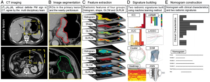
Occult peritoneal metastasis (PM) in advanced gastric cancer (AGC) patients is highly possible to be missed on computed tomography (CT) images. Patients with occult PMs are subject to late detection or even improper surgical treatment. We therefore aimed to develop a radiomic nomogram to preoperatively identify occult PMs in AGC patients.
A total of 554 AGC patients from 4 centers were divided into 1 training, 1 internal validation, and 2 external validation cohorts. All patients' PM status was firstly diagnosed as negative by CT, but later confirmed by laparoscopy (PM-positive n = 122, PM-negative n = 432). Radiomic signatures reflecting phenotypes of the primary tumor (RS1) and peritoneum region (RS2) were built as predictors of PM from 266 quantitative image features. Individualized nomograms of PM status incorporating RS1, RS2, or clinical factors were developed and evaluated regarding prediction ability.
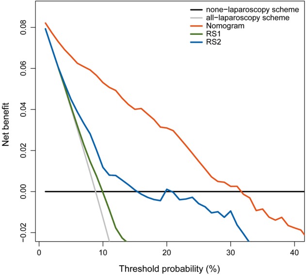
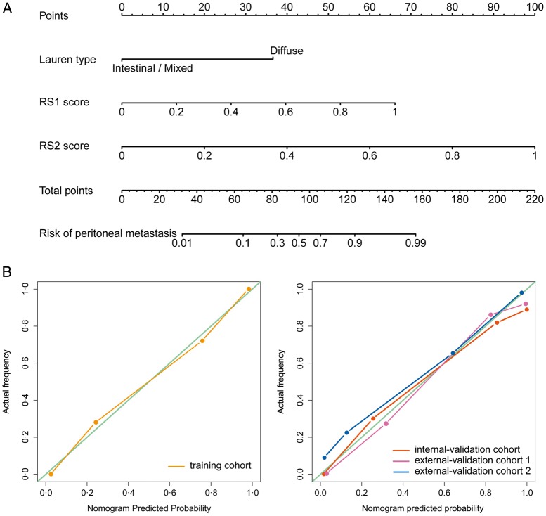
RS1, RS2, and Lauren type were significant predictors of occult PM (all P < 0.05). A nomogram of these three factors demonstrated better diagnostic accuracy than the model with RS1, RS2, or clinical factors alone (all net reclassification improvement P < 0.05). The area under curve yielded was 0.958 [95% confidence interval (CI) 0.923-0.993], 0.941 (95% CI 0.904-0.977), 0.928 (95% CI 0.886-0.971), and 0.920 (95% CI 0.862-0.978) for the training, internal, and two external validation cohorts, respectively. Stratification analysis showed that this nomogram had potential generalization ability.
CT phenotypes of both primary tumor and nearby peritoneum are significantly associated with occult PM status. A nomogram of these CT phenotypes and Lauren type has an excellent prediction ability of occult PM, and may have significant clinical implications on early detection of occult PM for AGC.
在晚期胃癌(AGC)患者中,计算机断层扫描(CT)图像上可能会高度漏诊隐匿性腹膜转移(PM)。隐匿性 PM 患者的检测往往较晚,甚至接受不适当的手术治疗。因此,我们旨在开发一种放射组学列线图,以术前识别 AGC 患者的隐匿性 PM。
来自 4 个中心的 554 名 AGC 患者被分为 1 个训练队列、1 个内部验证队列和 2 个外部验证队列。所有患者的 PM 状态首先通过 CT 诊断为阴性,但随后通过腹腔镜(PM 阳性 n=122,PM 阴性 n=432)确诊。从 266 个定量图像特征中构建反映原发肿瘤表型的放射组学特征(RS1)和腹膜区域的放射组学特征(RS2)作为 PM 的预测因子。分别建立并评估了纳入 RS1、RS2 或临床因素的个体化 PM 状态列线图的预测能力。
RS1、RS2 和 Lauren 型是隐匿性 PM 的显著预测因子(均 P<0.05)。这三个因素的列线图比仅包含 RS1、RS2 或临床因素的模型具有更好的诊断准确性(所有净重新分类改善 P<0.05)。训练、内部和两个外部验证队列的曲线下面积分别为 0.958(95%可信区间 0.923-0.993)、0.941(95%可信区间 0.904-0.977)、0.928(95%可信区间 0.886-0.971)和 0.920(95%可信区间 0.862-0.978)。分层分析表明,该列线图具有潜在的泛化能力。
原发肿瘤和附近腹膜的 CT 表型与隐匿性 PM 状态显著相关。该 CT 表型和 Lauren 型列线图对隐匿性 PM 具有极好的预测能力,可能对早期发现 AGC 的隐匿性 PM 具有重要的临床意义。