Singhal Mukesh Kumar, Dandriyal Ramakant, Aggarwal Asish, Agarwal Anshika, Yadav Sudhir, Baranwal Prashant
Department of Prosthodontics & Crown and Bridge Including Implantology, Institute of Dental Sciences, Bareilly, Uttar Pradesh, India.
Department of Oral Surgery, Institute of Dental Sciences, Bareilly, Uttar Pradesh, India.
Ann Maxillofac Surg. 2018 Jul-Dec;8(2):347-351. doi: 10.4103/ams.ams_161_17.
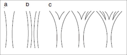
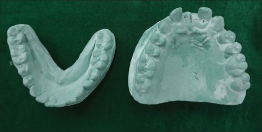
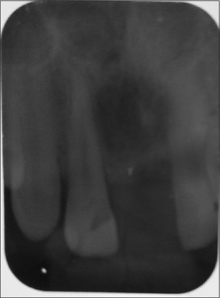
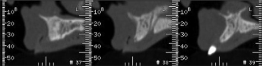
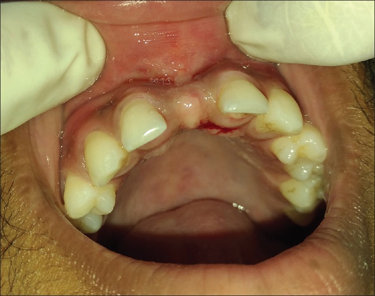
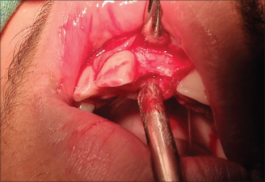
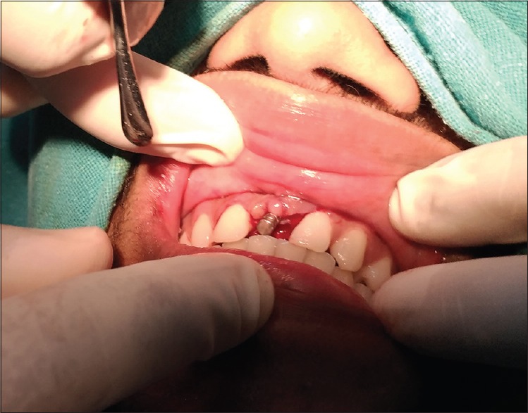
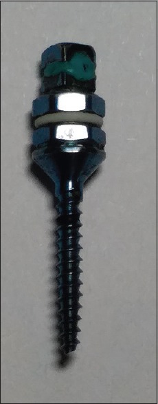
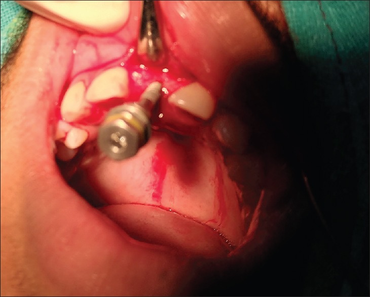
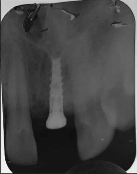
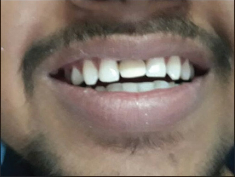
Implant placement is a challenge in the anterior maxilla if the available bone is reduced and esthetics is challenging. The ideal implant position should be considered in all three dimensions: mesiodistal, apicocoronal, and orofacial. This article includes a review and a case report for the anatomical and clinical perspective of implant placement in nasopalatine foramen (near incisal canal). In this case report, the edentulous space is mutilated in between the area #12 and #21 teeth. Therefore, only one, 3.0 W/10.00 L implant (bone size 4.2 mm width and 11 mm length) could be placed. Radiographically, D2 bone quality was diagnosed. Before surgery, an emphasis was given over the proper implant selection to avoid oversized implants due to critical anatomical landmark. Careful and with minimal trauma, the soft tissue was handled and implant placement was performed in a proper position, using information from panoramic radiograph, 3-D Dentascan. A surgical guide was used for placement of the implant. Finally, immediate loading of temporary implant prosthesis was done. The primary outcome was satisfactory, as after 72 h, no swelling and numbness were reported. The patient has been recalled after healing period of 24 weeks for permanent restoration.
如果上颌前部可用骨量减少且美学修复具有挑战性,种植体植入是一项挑战。理想的种植体位置应在三个维度上予以考虑:近远中向、根尖冠向和口面方向。本文从解剖学和临床角度对上颌腭中缝(切牙管附近)种植体植入进行综述并报告一例病例。在该病例报告中,12号和21号牙之间的无牙间隙已严重受损。因此,仅能植入一枚3.0W/10.00L种植体(骨尺寸为宽4.2mm、长11mm)。经影像学检查,诊断为D2级骨质量。手术前,着重强调了正确选择种植体,以避免因关键解剖标志而选用过大的种植体。借助全景X线片、三维牙科扫描提供的信息,轻柔且创伤最小地处理软组织,并将种植体植入合适位置。使用手术导板植入种植体。最后,即刻加载临时种植修复体。主要结果令人满意,术后72小时未报告肿胀和麻木情况。在24周的愈合期后召回患者进行永久修复。