Zhao Yuxue, Zhang Wenting, Jia Qi, Feng Zhendong, Guo Jing, Han Xueting, Liu Yuning, Shang Hongcai, Wang Yaoxian, Liu Wei Jing
Key Laboratory of Chinese Internal Medicine of Ministry of Education and Beijing, Dongzhimen Hospital Affiliated to Beijing University of Chinese Medicine, Beijing, China.
Renal Research Institution of Beijing University of Chinese Medicine, Dongzhimen Hospital Affiliated to Beijing University of Chinese Medicine, Beijing, China.
Front Physiol. 2019 Jan 21;9:1939. doi: 10.3389/fphys.2018.01939. eCollection 2018.
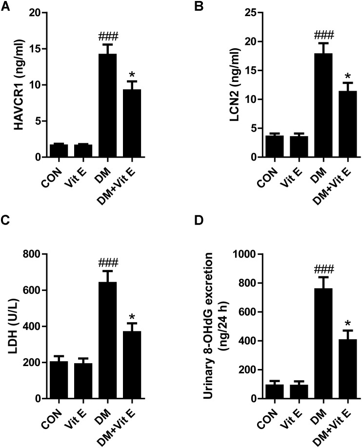
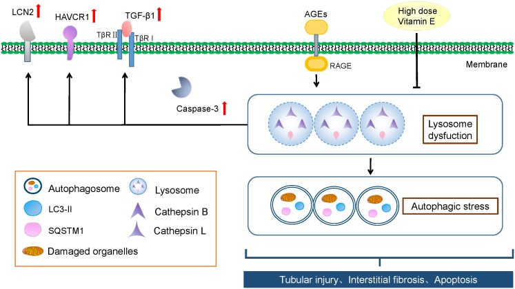
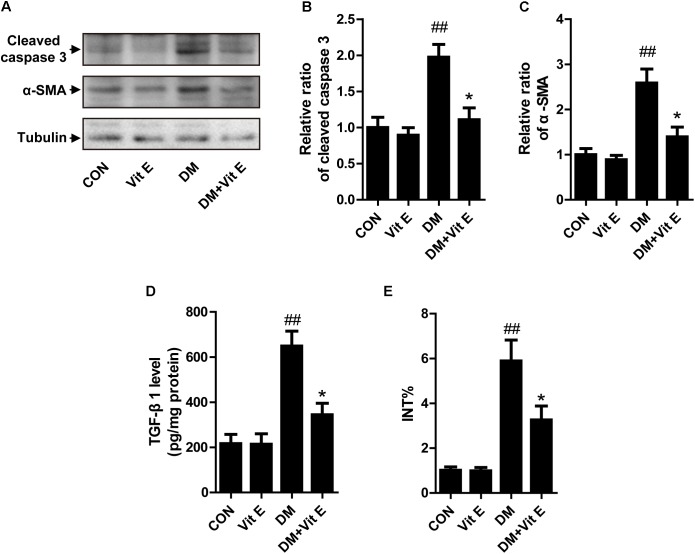
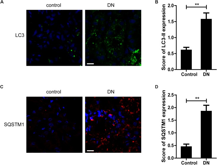
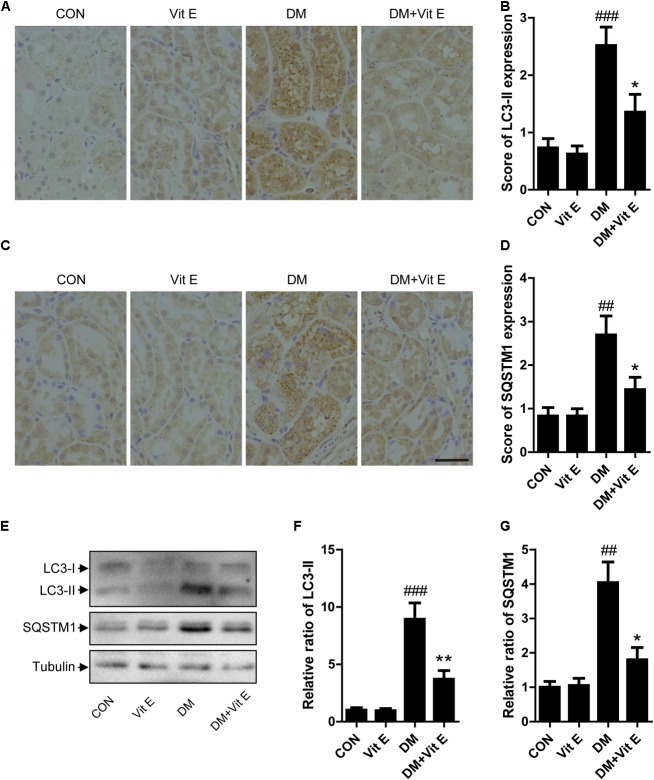
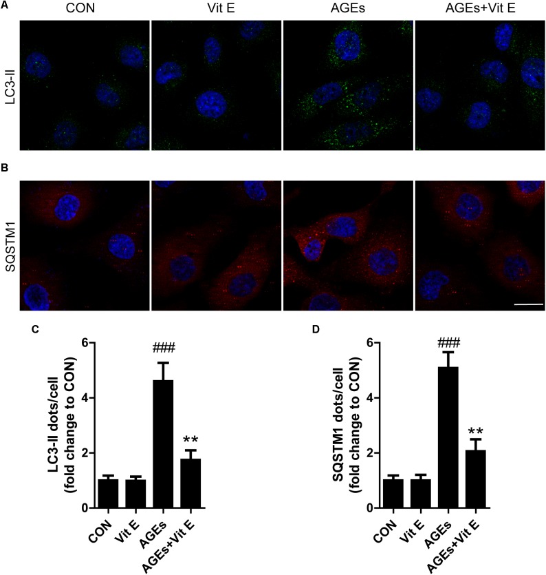
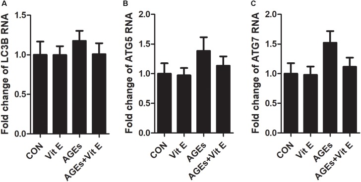
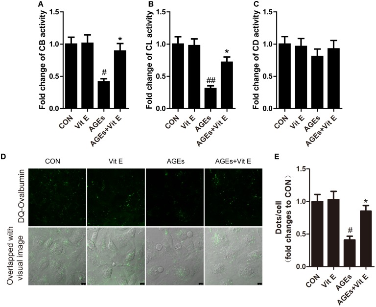
It has been reported that autophagic stress, which is involved in many diseases, plays a key role in the development of diabetic nephropathy (DN). In this study, we investigated the effects of high dose vitamin E on renal tubular epithelial cells and autophagic stress-related mechanisms in diabetes condition. In diabetic rats, high dose vitamin E treatment significantly decreased the serum creatinine, urea nitrogen, urinary albumin and urinary protein, reduced the levels of LCN2, HAVCR1, LDH and 8-OHdG in urine, and attenuated the cellular apoptosis and interstitial fibrosis in renal cortex. , vitamin E could reduce the release of LCN2 and HAVCR1 and the protein levels of caspase 3 and TGF-β1, as well as improve the growth inhibition in cultured HK-2 cells after exposure to advanced glycation end products (AGEs). Also, LC3-II and SQSTM1-positive dots were significantly increased in the renal tubular epithelial cells of DN patients and diabetic rats, and in HK-2 cells after exposure to AGEs, which were markedly declined by vitamin E. In addition, we found that the autophagosome formation was not affected by AGEs, as assessed by the mRNA levels of LC3B, Beclin-1, and ATG7. However, AGEs blocked the lysosomal degradation of autophagosome, which was characterized by a decrease in the enzymatic activity of cathepsin B/cathepsin L and DQ-ovalbumin degradation in HK-2 cells, indicating that AGEs-induced accumulation of autophagic vacuoles was a sign of autophagic stress. Interestingly, vitamin E exerted a protective effect on lysosomes to reduce the autophagic stress. Taken together, we conclude that autophagic stress may play an important part in the progression of DN, and alleviation of autophagic stress though improvement of lysosomal function provides a promising novel approach for treating DN.
据报道,自噬应激参与多种疾病,在糖尿病肾病(DN)的发展中起关键作用。在本研究中,我们研究了高剂量维生素E对糖尿病状态下肾小管上皮细胞和自噬应激相关机制的影响。在糖尿病大鼠中,高剂量维生素E治疗显著降低了血清肌酐、尿素氮、尿白蛋白和尿蛋白,降低了尿液中LCN2、HAVCR1、LDH和8-OHdG的水平,并减轻了肾皮质的细胞凋亡和间质纤维化。此外,维生素E可以减少LCN2和HAVCR1的释放以及半胱天冬酶3和TGF-β1的蛋白水平,并改善暴露于晚期糖基化终产物(AGEs)后培养的HK-2细胞中的生长抑制。而且,DN患者和糖尿病大鼠的肾小管上皮细胞以及暴露于AGEs后的HK-2细胞中LC3-II和SQSTM1阳性点显著增加,而维生素E使其明显减少。此外,我们发现自噬体的形成不受AGEs影响,通过LC3B、Beclin-1和ATG7的mRNA水平评估。然而,AGEs阻断了自噬体的溶酶体降解,其特征是HK-2细胞中组织蛋白酶B/组织蛋白酶L的酶活性降低和DQ-卵清蛋白降解减少,表明AGEs诱导的自噬泡积累是自噬应激的标志。有趣的是,维生素E对溶酶体发挥保护作用以减轻自噬应激。综上所述,我们得出结论,自噬应激可能在DN的进展中起重要作用,通过改善溶酶体功能减轻自噬应激为治疗DN提供了一种有前景的新方法。