Arrigo A P, Suhan J P, Welch W J
Cold Spring Harbor Laboratory, New York 11724.
Mol Cell Biol. 1988 Dec;8(12):5059-71. doi: 10.1128/mcb.8.12.5059-5071.1988.
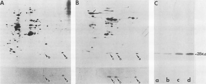
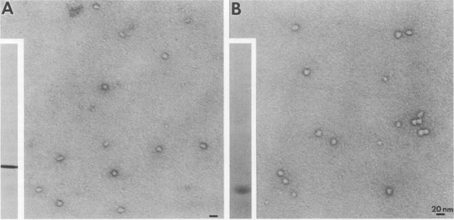
Mammalian cells grown at 37 degrees C contain a single low-molecular-weight heat shock (or stress) protein with an apparent mass of 28 kilodaltons (kDa) whose synthesis increases in cells after exposure to elevated temperatures or other forms of physiologic stress. Herein we present data demonstrating that heat shock protein 28 exists in a number of dynamic states depending upon the physiologic state of the cell. Biochemical fractionation of 37 degrees C cells in the absence of nonionic detergent revealed that the 28-kDa protein partitioned approximately equally between the soluble and insoluble fractions. The addition of detergent in the fractionation procedure resulted in all of the protein distributed within the soluble phase. In contrast, in cells first heat shocked and then fractionated in the presence of detergent, most of the 28-kDa protein was found within the insoluble fraction. These biochemical results appeared entirely consistent with indirect immunofluorescence experiments, demonstrating that the 28-kDa protein resided within the perinuclear region of 37 degrees C cells in close proximity to the Golgi complex. After heat shock treatment, the 28-kDa protein relocalized within the nucleus and resisted detergent extraction. The extent of 28-kDa protein redistribution into the nucleus and its detergent insolubility increased as a function of the severity of the heat shock treatment. With time of recovery from the heat treatment there occurred a gradual return of the 28-kDa protein into the detergent-soluble phase. Concomitant with these changes in 28-kDa protein solubility was a corresponding change in the apparent size of the protein as determined by gel filtration. While at 37 degrees C cells the protein exhibited a mass of 200 to 800 kDa; after heat shock the protein assumed sizes of 2 MDa or greater. Using immunoelectron microscopy, we show an accumulation of these aggregates of 28-kDa protein within the nucleus. Finally, we show that the heat-dependent redistribution of the 28-kDa protein from the cytoplasm into the nucleus was greatly diminished when the cells were first rendered thermotolerant, and we suggest that this simple assay (i.e., 28-kDa protein detergent solubility) may prove useful in evaluating the thermotolerant status of a cell or tissue.
在37摄氏度下培养的哺乳动物细胞含有一种单一的低分子量热休克(或应激)蛋白,其表观质量为28千道尔顿(kDa),在细胞暴露于高温或其他形式的生理应激后,其合成增加。在此我们提供的数据表明,热休克蛋白28根据细胞的生理状态以多种动态状态存在。在不存在非离子去污剂的情况下对37摄氏度的细胞进行生化分级分离,结果显示28-kDa蛋白在可溶部分和不溶部分中大致平均分配。在分级分离过程中加入去污剂后,所有蛋白质都分布在可溶相中。相反,在首先进行热休克然后在去污剂存在下进行分级分离的细胞中,大部分28-kDa蛋白存在于不溶部分中。这些生化结果似乎与间接免疫荧光实验完全一致,表明28-kDa蛋白位于37摄氏度细胞的核周区域,紧邻高尔基体复合体。热休克处理后,28-kDa蛋白重新定位于细胞核内并抵抗去污剂提取。28-kDa蛋白重新分布到细胞核中的程度及其去污剂不溶性随着热休克处理的严重程度而增加。随着从热处理中恢复的时间推移,28-kDa蛋白逐渐回到去污剂可溶相。与28-kDa蛋白溶解度的这些变化同时发生的是,通过凝胶过滤测定的蛋白质表观大小也相应改变。在37摄氏度的细胞中,该蛋白的质量为200至800 kDa;热休克后,该蛋白的大小为2 MDa或更大。使用免疫电子显微镜,我们显示这些28-kDa蛋白聚集体在细胞核内积累。最后,我们表明,当细胞首先被诱导产生耐热性时,28-kDa蛋白从细胞质到细胞核的热依赖性重新分布大大减少,并且我们认为这种简单的测定方法(即28-kDa蛋白去污剂溶解度)可能有助于评估细胞或组织的耐热状态。