Lister Hill National Center for Biomedical Communications, Communications Engineering Branch, National Library of Medicine, National Institutes of Health, Bethesda, USA.
Int J Comput Assist Radiol Surg. 2019 Apr;14(4):563-576. doi: 10.1007/s11548-019-01917-1. Epub 2019 Feb 7.
Chest radiography is the most common imaging modality for pulmonary diseases. Due to its wide usage, there is a rich literature addressing automated detection of cardiopulmonary diseases in digital chest X-rays (CXRs). One of the essential steps for automated analysis of CXRs is localizing the relevant region of interest, i.e., isolating lung region from other less relevant parts, for applying decision-making algorithms there. This article provides an overview of the recent literature on lung boundary detection in CXR images.
We review the leading lung segmentation algorithms proposed in period 2006-2017. First, we present a review of articles for posterior-anterior view CXRs. Then, we mention studies which operate on lateral views. We pay particular attention to works that focus their efforts on deformed lungs and pediatric cases. We also highlight the radiographic measures extracted from lung boundary and their use in automatically detecting cardiopulmonary abnormalities. Finally, we identify challenges in dataset curation and expert delineation process, and we listed publicly available CXR datasets.
(1) We classified algorithms into four categories: rule-based, pixel classification-based, model-based, hybrid, and deep learning-based algorithms. Based on the reviewed articles, hybrid methods and deep learning-based methods surpass the algorithms in other classes and have segmentation performance as good as inter-observer performance. However, they require long training process and pose high computational complexity. (2) We found that most of the algorithms in the literature are evaluated on posterior-anterior view adult CXRs with a healthy lung anatomy appearance without considering challenges in abnormal CXRs. (3) We also found that there are limited studies for pediatric CXRs. The lung appearance in pediatrics, especially in infant cases, deviates from adult lung appearance due to the pediatric development stages. Moreover, pediatric CXRs are noisier than adult CXRs due to interference by other objects, such as someone holding the child's arms or the child's body, and irregular body pose. Therefore, lung boundary detection algorithms developed on adult CXRs may not perform accurately in pediatric cases and need additional constraints suitable for pediatric CXR imaging characteristics. (4) We have also stated that one of the main challenges in medical image analysis is accessing the suitable datasets. We listed benchmark CXR datasets for developing and evaluating the lung boundary algorithms. However, the number of CXR images with reference boundaries is limited due to the cumbersome but necessary process of expert boundary delineation.
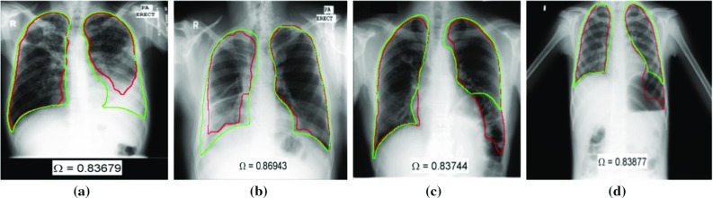
A reliable computer-aided diagnosis system would need to support a greater variety of lung and background appearance. To our knowledge, algorithms in the literature are evaluated on posterior-anterior view adult CXRs with a healthy lung anatomy appearance, without considering ambiguous lung silhouettes due to pathological deformities, anatomical alterations due to misaligned body positioning, patient's development stage and gross background noises such as holding hands, jewelry, patient's head and legs in CXR. Considering all the challenges which are not very well addressed in the literature, developing lung boundary detection algorithms that are robust to such interference remains a challenging task. We believe that a broad review of lung region detection algorithms would be useful for researchers working in the field of automated detection/diagnosis algorithms for lung/heart pathologies in CXRs.

胸部 X 射线摄影是肺部疾病最常用的影像学检查方法。由于其广泛的应用,有大量文献研究数字胸部 X 射线(CXR)中心肺疾病的自动检测。CXR 自动分析的一个基本步骤是定位相关的感兴趣区域,即从其他不相关的部分隔离肺区域,以便在那里应用决策算法。本文综述了 2006 年至 2017 年期间有关 CXR 图像中肺边界检测的最新文献。
我们回顾了 2006 年至 2017 年期间提出的主要肺分割算法。首先,我们对后前位 CXR 的文章进行了综述。然后,我们提到了适用于侧位 CXR 的研究。我们特别关注那些专注于变形肺和儿科病例的研究。我们还强调了从肺边界提取的放射学指标及其在自动检测心肺异常中的应用。最后,我们确定了数据集整理和专家勾画过程中的挑战,并列出了公开的 CXR 数据集。
(1)我们将算法分为四类:基于规则、基于像素分类、基于模型、混合和基于深度学习的算法。根据综述的文章,混合方法和基于深度学习的方法优于其他类别的算法,具有与观察者间性能相当的分割性能。然而,它们需要较长的训练过程,并且具有较高的计算复杂度。(2)我们发现,文献中的大多数算法都是在后前位成人 CXR 上进行评估的,这些 CXR 具有健康的肺解剖结构,而没有考虑异常 CXR 中的挑战。(3)我们还发现,儿科 CXR 的研究有限。儿科,尤其是婴儿期,由于儿童发育阶段的原因,肺的外观与成人肺的外观不同。此外,由于其他物体(如抱着孩子的手臂或孩子的身体)的干扰以及不规则的身体姿势,儿科 CXR 比成人 CXR 噪声更大。因此,在成人 CXR 上开发的肺边界检测算法可能在儿科病例中无法准确执行,需要额外的适合儿科 CXR 成像特征的约束条件。(4)我们还指出,医学图像分析中的主要挑战之一是访问合适的数据集。我们列出了用于开发和评估肺边界算法的基准 CXR 数据集。然而,由于专家边界勾画过程繁琐但必要,具有参考边界的 CXR 图像数量有限。
一个可靠的计算机辅助诊断系统需要支持更多种类的肺和背景外观。据我们所知,文献中的算法是在后前位成人 CXR 上进行评估的,这些 CXR 具有健康的肺解剖结构,没有考虑到由于病理性变形、由于身体定位不当引起的解剖改变、患者发育阶段以及 CXR 中的手部、珠宝、患者头部和腿部等大体背景噪声引起的模糊肺轮廓。考虑到文献中没有很好解决的所有挑战,开发对这些干扰具有鲁棒性的肺边界检测算法仍然是一项具有挑战性的任务。我们相信,对肺区域检测算法的广泛综述将对从事肺/心脏疾病在 CXR 中自动检测/诊断算法研究的研究人员有用。