Department of Dermatology, Complejo Hospitalario de Toledo, Toledo, Spain.
Department of Radiology, Gregorio Marañon Hospital, Madrid, Spain.
PLoS One. 2019 Feb 8;14(2):e0211808. doi: 10.1371/journal.pone.0211808. eCollection 2019.
Psoriasis is associated with an increased risk of cardiovascular disease (CVD) at younger ages that is not identifiable by traditional risk factors. Screening for subclinical atherosclerosis with ultrasound has only been investigated in carotid arteries. Femoral artery ultrasound has never been considered for this purpose. The link between psoriasis and accelerated atherosclerosis has not yet been established.
To study the usefulness of femoral artery ultrasound for the detection of subclinical atherosclerosis in psoriasis. We also investigated its possible relationship with changes in insulin resistance.
We conducted a cross-sectional study in 140 participants, 70 patients with moderate-to-severe psoriasis and 70 healthy controls, matched 1:1 for age, sex, and BMI. Femoral and carotid atherosclerotic plaques were evaluated by ultrasonography. Insulin resistance was assessed by the homeostasis model assessment method (HOMA-IR).
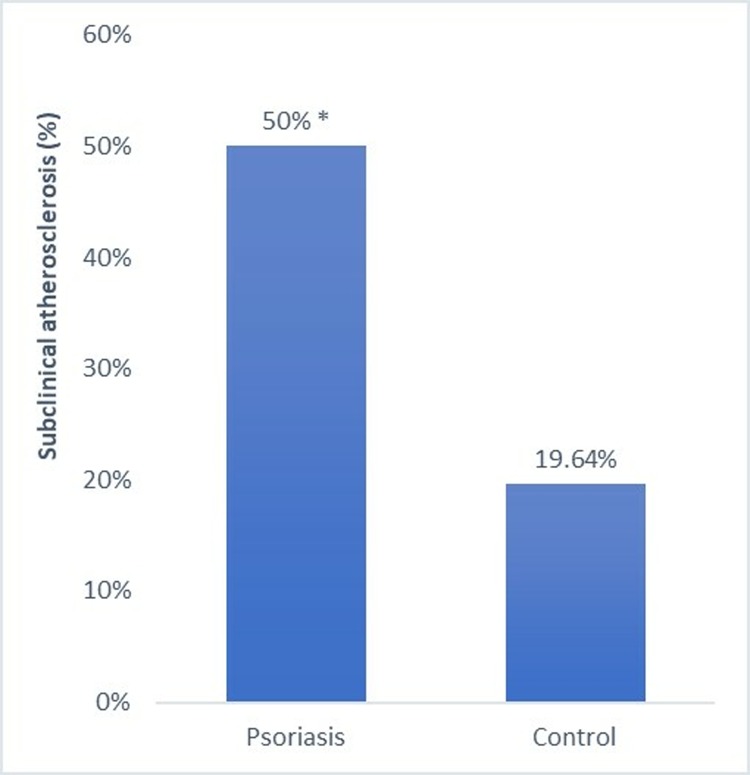
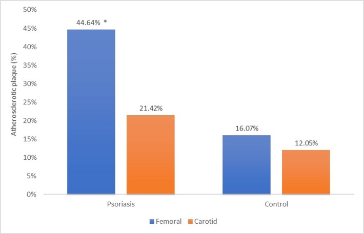
Femoral atherosclerotic plaque prevalence was significantly higher in patients with psoriasis (44.64%) than in controls (19.07%) (p<0.005), but no significant difference was found in carotid plaque prevalence (p<0.3). Femoral plaques were significantly more prevalent than carotid plaques (21.42%) among patients with psoriasis (p<0.001). In the regression analysis, insulin resistance was the most influential determinant of atherosclerosis in psoriasis and C-reactive protein the most significant predictor of insulin resistance.
Ultrasound screening for femoral atherosclerotic plaques improves the detection of subclinical atherosclerosis in patients with psoriasis, whereas the study of carotid arteries is not sufficiently accurate. Insulin resistance appears to play a greater role in the development of atherosclerosis in these patients in comparison to other classical CVD risk factors.
银屑病患者发生心血管疾病(CVD)的风险会随年龄增长而增加,且这种风险无法通过传统的危险因素进行识别。使用超声检查颈动脉粥样硬化的亚临床病变仅在颈动脉中进行了研究。从未考虑过使用股动脉超声进行这种检查。银屑病与动脉粥样硬化加速之间的联系尚未得到证实。
研究股动脉超声在检测银屑病患者亚临床动脉粥样硬化中的作用。我们还研究了它与胰岛素抵抗变化之间的可能关系。
我们进行了一项横断面研究,共纳入 140 名参与者,其中 70 名患者为中重度银屑病,70 名为健康对照,按年龄、性别和 BMI 1:1 匹配。通过超声检查评估股动脉和颈动脉粥样硬化斑块。采用稳态模型评估法(HOMA-IR)评估胰岛素抵抗。
银屑病患者的股动脉粥样硬化斑块发生率(44.64%)明显高于对照组(19.07%)(p<0.005),但颈动脉斑块发生率无显著差异(p<0.3)。银屑病患者的股动脉斑块发生率(21.42%)明显高于颈动脉斑块发生率(p<0.001)。在回归分析中,胰岛素抵抗是银屑病患者动脉粥样硬化的最主要影响因素,C 反应蛋白是胰岛素抵抗的最主要预测因子。
超声筛查股动脉粥样硬化斑块可提高对银屑病患者亚临床动脉粥样硬化的检出率,而研究颈动脉则不够准确。与其他经典 CVD 危险因素相比,胰岛素抵抗似乎在这些患者的动脉粥样硬化发展中起更大作用。