Department of Radiology, Johns Hopkins University School of Medicine, Baltimore, MD, USA; F.M. Kirby Research Center for Functional Brain Imaging, Kennedy Krieger Institute, Baltimore, MD, USA.
Department of Radiology, Johns Hopkins University School of Medicine, Baltimore, MD, USA; F.M. Kirby Research Center for Functional Brain Imaging, Kennedy Krieger Institute, Baltimore, MD, USA; Department of Electronic Science, Fujian Provincial Key Laboratory of Plasma and Magnetic Resonance, Xiamen University, Xiamen, China.
Neuroimage. 2019 May 1;191:337-349. doi: 10.1016/j.neuroimage.2019.02.016. Epub 2019 Feb 7.
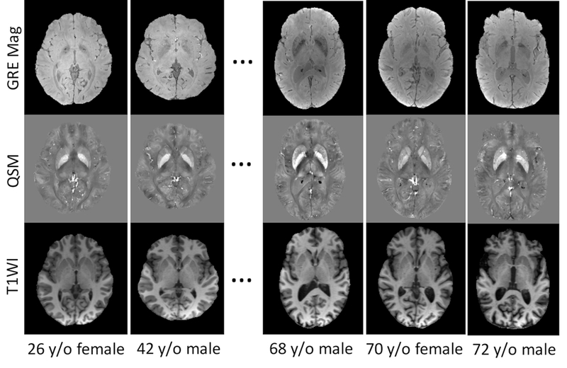
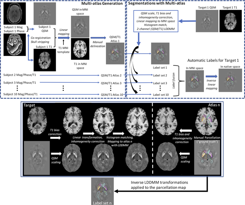
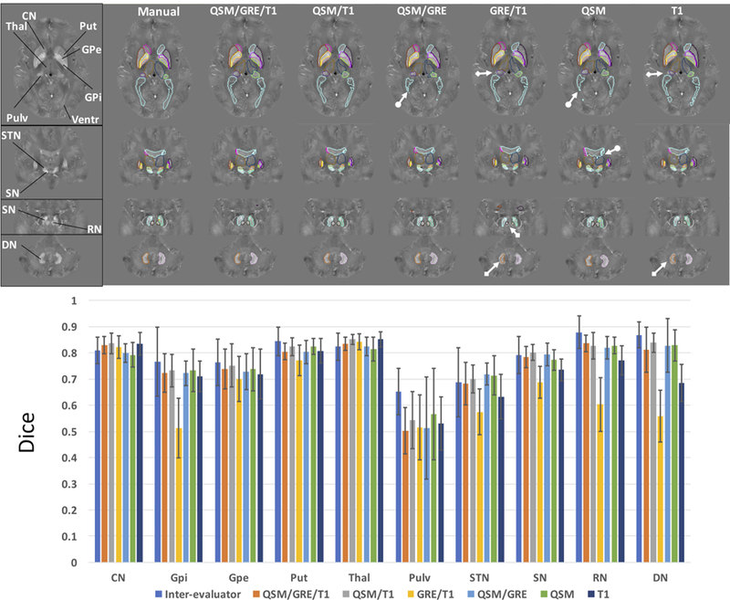
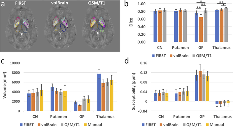
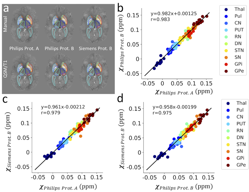
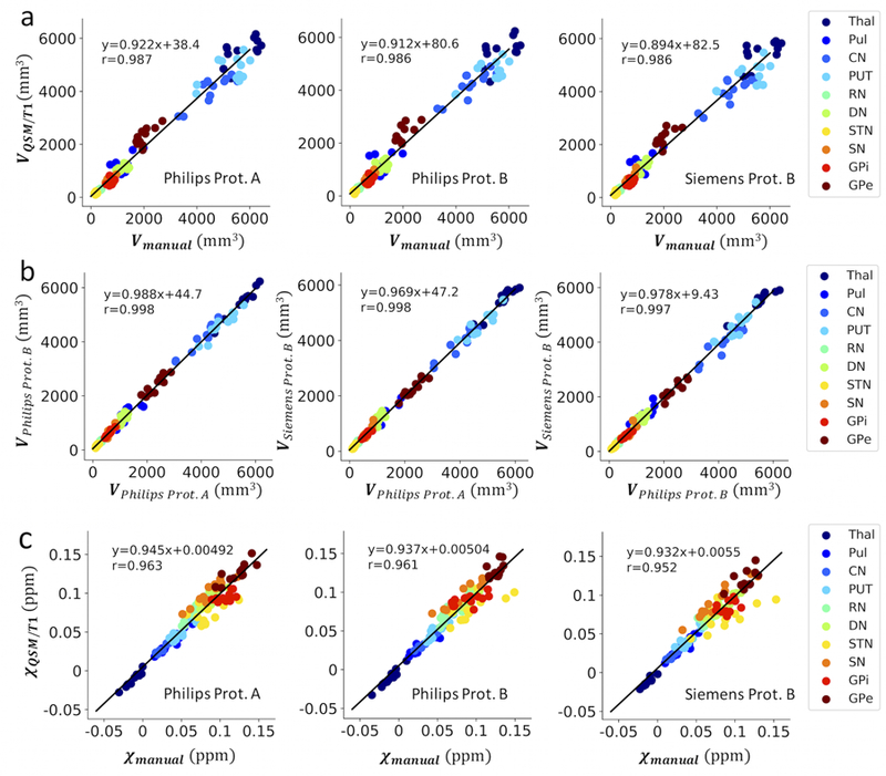
Quantification of tissue magnetic susceptibility using MRI offers a non-invasive measure of important tissue components in the brain, such as iron and myelin, potentially providing valuable information about normal and pathological conditions during aging. Despite many advances made in recent years on imaging techniques of quantitative susceptibility mapping (QSM), accurate and robust automated segmentation tools for QSM images that can help generate universal and sharable susceptibility measures in a biologically meaningful set of structures are still not widely available. In the present study, we developed an automated process to segment brain nuclei and quantify tissue susceptibility in these regions based on a susceptibility multi-atlas library, consisting of 10 atlases with T1-weighted images, gradient echo (GRE) magnitude images and QSM images of brains with different anatomic patterns. For each atlas in this library, 10 regions of interest in iron-rich deep gray matter structures that are better defined by QSM contrast were manually labeled, including caudate, putamen, globus pallidus internal/external, thalamus, pulvinar, subthalamic nucleus, substantia nigra, red nucleus and dentate nucleus in both left and right hemispheres. We then tested different pipelines using different combinations of contrast channels to bring the set of labels from the multi-atlases to each target brain and compared them with the gold standard manual delineation. The results showed that the segmentation accuracy using dual contrasts QSM/T1 pipeline outperformed other dual-contrast or single-contrast pipelines. The dice values of 0.77 ± 0.09 using the QSM/T1 multi-atlas pipeline rivaled with the segmentation reliability obtained from multiple evaluators with dice values of 0.79 ± 0.07 and gave comparable or superior performance in segmenting subcortical nuclei in comparison with standard FSL FIRST or recent multi-atlas package of volBrain. The segmentation performance of the QSM/T1 multi-atlas was further tested on QSM images acquired using different acquisition protocols and platforms and showed good reliability and reproducibility with average dice of 0.79 ± 0.08 to manual labels and 0.89 ± 0.04 in an inter-protocol manner. The extracted quantitative magnetic susceptibility values in the deep gray matter nuclei also correlated well between different protocols with inter-protocol correlation constants all larger than 0.97. Such reliability and performance was ultimately validated in an external dataset acquired at another study site with consistent susceptibility measures obtained using the QSM/T1 multi-atlas approach in comparison to those using manual delineation. In summary, we designed a susceptibility multi-atlas tool for automated and reliable segmentation of QSM images and for quantification of magnetic susceptibilities. It is publicly available through our cloud-based platform (www.mricloud.org). Further improvement on the performance of this multi-atlas tool is expected by increasing the number of atlases in the future.
利用 MRI 对组织磁化率进行定量分析,可以提供脑内重要组织成分(如铁和髓鞘)的非侵入性测量,这可能为衰老过程中的正常和病理状态提供有价值的信息。尽管近年来在定量磁化率映射(QSM)的成像技术方面取得了许多进展,但对于 QSM 图像,仍然缺乏能够在一组具有生物学意义的结构中生成通用且可共享的磁化率测量值的准确、稳健的自动化分割工具。在本研究中,我们开发了一种基于磁化率多图谱库的自动处理流程,用于对脑核进行分割和量化这些区域的组织磁化率。该多图谱库由 10 个图谱组成,每个图谱都包含 T1 加权图像、梯度回波(GRE)幅度图像和具有不同解剖模式的大脑的 QSM 图像。对于该库中的每个图谱,手动标记了 10 个富含铁的深部灰质结构中的感兴趣区域(ROI),这些 ROI 在 QSM 对比度的定义上更好,包括左右半球的尾状核、壳核、苍白球内外侧、丘脑、丘脑下核、黑质、红核和齿状核。然后,我们使用不同的对比度通道组合测试了不同的管道,将多图谱中的标签集带到每个目标大脑,并将其与金标准手动勾画进行比较。结果表明,使用双对比度 QSM/T1 管道的分割准确性优于其他双对比度或单对比度管道。使用 QSM/T1 多图谱管道获得的 0.77±0.09 的 Dice 值与多位评估者获得的 0.79±0.07 的分割可靠性相当,并在与标准 FSL FIRST 或最近的 volBrain 多图谱包相比时,在分割皮质下核方面具有可比或更好的性能。QSM/T1 多图谱的分割性能还在使用不同采集协议和平台采集的 QSM 图像上进行了测试,显示出良好的可靠性和可重复性,与手动标签的平均 Dice 值为 0.79±0.08,在协议间方式下为 0.89±0.04。在不同协议之间,深部灰质核中的定量磁化率值也具有很好的相关性,协议间相关常数均大于 0.97。通过使用 QSM/T1 多图谱方法与手动勾画方法相比,在另一个研究地点获得的外部数据集上最终验证了这种可靠性和性能,得到了一致的磁化率测量值。总之,我们设计了一种用于 QSM 图像自动和可靠分割以及磁化率定量分析的磁化率多图谱工具。它可通过我们的云平台(www.mricloud.org)公开获取。通过增加图谱的数量,预计该多图谱工具的性能将进一步提高。