Herynek Vít, Turnovcová Karolína, Gálisová Andrea, Kaman Ondřej, Mareková Dana, Koktan Jakub, Vosmanská Magda, Kosinová Lucie, Jendelová Pavla
Radiodiagnostic and Interventional Radiology Department Institute for Clinical and Experimental Medicine Vídeňská 1958/9 140 21 Prague Czech Republic.
Center for Advanced Preclinical Imaging First Faculty of Medicine Charles University Salmovská 3 Prague Czech Republic.
ChemistryOpen. 2019 Jan 23;8(2):155-165. doi: 10.1002/open.201800261. eCollection 2019 Feb.
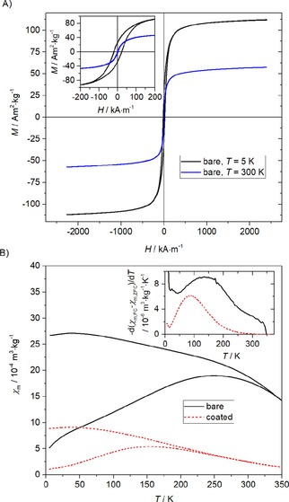
Manganese-zinc ferrite nanoparticles were synthesized by using a hydrothermal treatment, coated with silica, and then tested as efficient cellular labels for cell tracking, using magnetic resonance imaging (MRI) in vivo. A toxicity study was performed on rat mesenchymal stem cells and C6 glioblastoma cells. Adverse effects on viability and cell proliferation were observed at the highest concentration (0.55 mM) only; cell viability was not compromised at lower concentrations. Nanoparticle internalization was confirmed by transmission electron microscopy. The particles were found in membranous vesicles inside the cytoplasm. Although the metal content (0.42 pg Fe/cell) was lower compared to commercially available iron oxide nanoparticles, labeled cells reached a comparable relaxation rate , owing to higher nanoparticle relaxivity. Cells from transgenic luciferase-positive rats were used for in vivo experiments. Labeled cells were transplanted into the muscles of non-bioluminescent rats and visualized by MRI. The cells produced a distinct hypointense signal in T- or T*-weighted MR images in vivo. Cell viability in vivo was verified by bioluminescence.
通过水热法合成了锰锌铁氧体纳米颗粒,并用二氧化硅进行包覆,然后将其作为高效的细胞标记物用于体内细胞追踪的磁共振成像(MRI)检测。对大鼠间充质干细胞和C6胶质母细胞瘤细胞进行了毒性研究。仅在最高浓度(0.55 mM)时观察到对细胞活力和增殖的不利影响;较低浓度下细胞活力未受影响。通过透射电子显微镜确认了纳米颗粒的内化。在细胞质内的膜泡中发现了这些颗粒。尽管与市售的氧化铁纳米颗粒相比,金属含量(0.42 pg Fe/细胞)较低,但由于纳米颗粒具有更高的弛豫率,标记细胞达到了相当的弛豫率。来自转基因荧光素酶阳性大鼠的细胞用于体内实验。将标记的细胞移植到非生物发光大鼠的肌肉中,并通过MRI进行可视化。在体内的T加权或T*加权MR图像中,这些细胞产生了明显的低信号。通过生物发光验证了体内细胞的活力。