Danish Dementia Research Centre, Department of Neurology, Rigshospitalet, University of Copenhagen, Denmark.
Combinostics Ltd., Tampere, Finland.
Neuroimage Clin. 2019;22:101711. doi: 10.1016/j.nicl.2019.101711. Epub 2019 Feb 4.
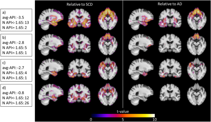
Diagnosing frontotemporal dementia may be challenging. New methods for analysis of regional brain atrophy patterns on magnetic resonance imaging (MRI) could add to the diagnostic assessment. Therefore, we aimed to develop automated imaging biomarkers for differentiating frontotemporal dementia subtypes from other diagnostic groups, and from one another.
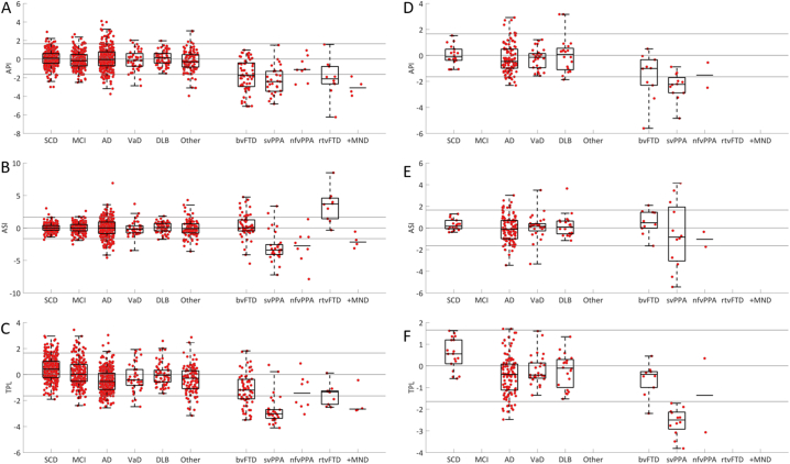
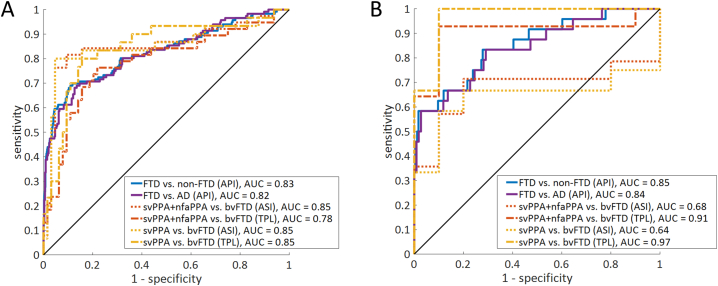
In this retrospective multicenter cohort study, we included 1213 patients (age 67 ± 9, 48% females) from two memory clinic cohorts: 116 frontotemporal dementia, 341 Alzheimer's disease, 66 Dementia with Lewy bodies, 40 vascular dementia, 104 other dementias, 229 mild cognitive impairment, and 317 subjective cognitive decline. Three MRI atrophy biomarkers were derived from the normalized volumes of automatically segmented cortical regions: 1) the anterior vs. posterior index, 2) the asymmetry index, and 3) the temporal pole left index. We used the following performance metrics: area under the receiver operating characteristic curve (AUC), sensitivity, and specificity. To account for the low prevalence of frontotemporal dementia we pursued a high specificity of 95%. Cross-validation was used in assessing the performance. The generalizability was assessed in an independent cohort (n = 200).
The anterior vs. posterior index performed with an AUC of 83% for differentiation of frontotemporal dementia from all other diagnostic groups (Sensitivity = 59%, Specificity = 95%, positive likelihood ratio = 11.8, negative likelihood ratio = 0.4). The asymmetry index showed highest performance for separation of primary progressive aphasia and behavioral variant frontotemporal dementia (AUC = 85%, Sensitivity = 79%, Specificity = 92%, positive likelihood ratio = 9.9, negative likelihood ratio = 0.2), whereas the temporal pole left index was specific for detection of semantic variant primary progressive aphasia (AUC = 85%, Sensitivity = 82%, Specificity = 80%, positive likelihood ratio = 4.1, negative likelihood ratio = 0.2). The validation cohort provided corresponding results for the anterior vs. posterior index and temporal pole left index.
This study presents three quantitative MRI biomarkers, which could provide additional information to the diagnostic assessment and assist clinicians in diagnosing frontotemporal dementia.
诊断额颞叶痴呆可能具有挑战性。磁共振成像(MRI)上区域性脑萎缩模式的新分析方法可以辅助诊断评估。因此,我们旨在开发自动成像生物标志物,以区分额颞叶痴呆亚型与其他诊断组以及彼此之间的区别。
在这项回顾性多中心队列研究中,我们纳入了来自两个记忆诊所队列的 1213 名患者(年龄 67±9 岁,48%为女性):116 名额颞叶痴呆患者、341 名阿尔茨海默病患者、66 名路易体痴呆患者、40 名血管性痴呆患者、104 名其他痴呆患者、229 名轻度认知障碍患者和 317 名主观认知减退患者。从自动分割的皮质区域的归一化体积中得出了三个 MRI 萎缩生物标志物:1)前/后指数,2)不对称指数,和 3)左侧颞极指数。我们使用以下性能指标:接收者操作特征曲线下的面积(AUC)、敏感度和特异性。为了考虑到额颞叶痴呆的低患病率,我们追求 95%的高特异性。交叉验证用于评估性能。在一个独立的队列(n=200)中评估了泛化能力。
前/后指数用于区分额颞叶痴呆与所有其他诊断组的 AUC 为 83%(敏感度=59%,特异性=95%,阳性似然比=11.8,阴性似然比=0.4)。不对称指数在区分原发性进行性失语症和行为变异型额颞叶痴呆方面表现出最高的性能(AUC=85%,敏感度=79%,特异性=92%,阳性似然比=9.9,阴性似然比=0.2),而左侧颞极指数则特异性地检测语义变异型原发性进行性失语症(AUC=85%,敏感度=82%,特异性=80%,阳性似然比=4.1,阴性似然比=0.2)。验证队列为前/后指数和左侧颞极指数提供了相应的结果。
本研究提出了三个定量 MRI 生物标志物,可为诊断评估提供额外信息,并协助临床医生诊断额颞叶痴呆。