• 文献检索
  • 文档翻译
  • 深度研究
  • 学术资讯
  • Suppr Zotero 插件Zotero 插件
  • 邀请有礼
  • 套餐&价格
  • 历史记录
应用&插件
Suppr Zotero 插件Zotero 插件浏览器插件Mac 客户端Windows 客户端微信小程序
定价
高级版会员购买积分包购买API积分包
服务
文献检索文档翻译深度研究API 文档MCP 服务
关于我们
关于 Suppr公司介绍联系我们用户协议隐私条款
关注我们

Suppr 超能文献

核心技术专利:CN118964589B侵权必究
粤ICP备2023148730 号-1Suppr @ 2026

文献检索

告别复杂PubMed语法,用中文像聊天一样搜索,搜遍4000万医学文献。AI智能推荐,让科研检索更轻松。

立即免费搜索

文件翻译

保留排版,准确专业,支持PDF/Word/PPT等文件格式,支持 12+语言互译。

免费翻译文档

深度研究

AI帮你快速写综述,25分钟生成高质量综述,智能提取关键信息,辅助科研写作。

立即免费体验

基于 d-Eye 传感器和图像处理算法的远程医疗系统自动检测视网膜彩色图像中硬性渗出物的方法。

An Approach to Automatic Hard Exudate Detection in Retina Color Images by a Telemedicine System Based on the d-Eye Sensor and Image Processing Algorithms.

机构信息

Department of Ophthalmology, Faculty of Medicine, Medical University of Bialystok, 24A Curie-Sklodowskiej Street, 15-276 Bialystok, Poland.

Bialystok University of Technology, Faculty of Computer Science, 45A Wiejska Street,15-351 Białystok, Poland.

出版信息

Sensors (Basel). 2019 Feb 8;19(3):695. doi: 10.3390/s19030695.

DOI:10.3390/s19030695
PMID:30744032
原文链接:https://pmc.ncbi.nlm.nih.gov/articles/PMC6387053/
Abstract

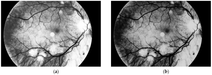
Hard exudates are one of the most characteristic and dangerous signs of diabetic retinopathy. They can be marked during the routine ophthalmological examination and seen in color fundus photographs (i.e., using a fundus camera). The purpose of this paper is to introduce an algorithm that can extract pathological changes (i.e., hard exudates) in diabetic retinopathy. This was a retrospective, nonrandomized study. A total of 100 photos were included in the analysis-50 sick and 50 normal eyes. Small lesions in diabetic retinopathy could be automatically diagnosed by the system with an accuracy of 98%. During the experiments, the authors used classical image processing methods such as binarization or median filtration, and data was read from the d-Eye sensor. Sixty-seven patients (39 females and 28 males with ages ranging between 50 and 64) were examined. The results have shown that the proposed solution accuracy level equals 98%. Moreover, the algorithm returns correct classification decisions for high quality images and low quality samples. Furthermore, we consider taking retina photos using mobile phones rather than fundus cameras, which is more practical. The paper presents an innovative approach. The results are introduced and the algorithm is described.

摘要

硬性渗出物是糖尿病视网膜病变最具特征性和危险性的标志之一。它们可以在常规眼科检查中被标记,并在眼底彩色照片中看到(即使用眼底相机)。本文的目的是介绍一种可以提取糖尿病性视网膜病变中病理性改变(即硬性渗出物)的算法。这是一项回顾性、非随机研究。分析中总共包括 100 张照片,其中 50 张是患病眼睛的照片,50 张是正常眼睛的照片。该系统可以自动诊断糖尿病视网膜病变中的小病变,准确率达到 98%。在实验过程中,作者使用了经典的图像处理方法,如二值化或中值滤波,并从 d-Eye 传感器读取数据。共检查了 67 名患者(39 名女性和 28 名男性,年龄在 50 至 64 岁之间)。结果表明,所提出的解决方案的准确率达到 98%。此外,该算法对高质量图像和低质量样本都能返回正确的分类决策。此外,我们还考虑使用手机而不是眼底相机拍摄视网膜照片,这更实用。本文提出了一种创新的方法。介绍了结果并描述了算法。

https://cdn.ncbi.nlm.nih.gov/pmc/blobs/ebf8/6387053/783df0628855/sensors-19-00695-g019.jpg
https://cdn.ncbi.nlm.nih.gov/pmc/blobs/ebf8/6387053/a9fb8c941241/sensors-19-00695-g001.jpg
https://cdn.ncbi.nlm.nih.gov/pmc/blobs/ebf8/6387053/45456edf7af2/sensors-19-00695-g002a.jpg
https://cdn.ncbi.nlm.nih.gov/pmc/blobs/ebf8/6387053/53c0b492aeec/sensors-19-00695-g003.jpg
https://cdn.ncbi.nlm.nih.gov/pmc/blobs/ebf8/6387053/d0e642fcdf06/sensors-19-00695-g004.jpg
https://cdn.ncbi.nlm.nih.gov/pmc/blobs/ebf8/6387053/d06e4a6ce183/sensors-19-00695-g005.jpg
https://cdn.ncbi.nlm.nih.gov/pmc/blobs/ebf8/6387053/200c01b92b96/sensors-19-00695-g006.jpg
https://cdn.ncbi.nlm.nih.gov/pmc/blobs/ebf8/6387053/e64c76fae8fc/sensors-19-00695-g007.jpg
https://cdn.ncbi.nlm.nih.gov/pmc/blobs/ebf8/6387053/fdfbd7aaeb5d/sensors-19-00695-g008.jpg
https://cdn.ncbi.nlm.nih.gov/pmc/blobs/ebf8/6387053/11c89844d947/sensors-19-00695-g009.jpg
https://cdn.ncbi.nlm.nih.gov/pmc/blobs/ebf8/6387053/eb971382ccda/sensors-19-00695-g010.jpg
https://cdn.ncbi.nlm.nih.gov/pmc/blobs/ebf8/6387053/76c053273835/sensors-19-00695-g011.jpg
https://cdn.ncbi.nlm.nih.gov/pmc/blobs/ebf8/6387053/7ea4a9f75b35/sensors-19-00695-g012.jpg
https://cdn.ncbi.nlm.nih.gov/pmc/blobs/ebf8/6387053/2f1bd000e5b6/sensors-19-00695-g013.jpg
https://cdn.ncbi.nlm.nih.gov/pmc/blobs/ebf8/6387053/fa5de4838c06/sensors-19-00695-g014.jpg
https://cdn.ncbi.nlm.nih.gov/pmc/blobs/ebf8/6387053/5b1e17465d4d/sensors-19-00695-g015.jpg
https://cdn.ncbi.nlm.nih.gov/pmc/blobs/ebf8/6387053/8c6af13b1580/sensors-19-00695-g016.jpg
https://cdn.ncbi.nlm.nih.gov/pmc/blobs/ebf8/6387053/08ca11ba4d63/sensors-19-00695-g017.jpg
https://cdn.ncbi.nlm.nih.gov/pmc/blobs/ebf8/6387053/5a19c475d7be/sensors-19-00695-g018.jpg
https://cdn.ncbi.nlm.nih.gov/pmc/blobs/ebf8/6387053/783df0628855/sensors-19-00695-g019.jpg

相似文献

1
An Approach to Automatic Hard Exudate Detection in Retina Color Images by a Telemedicine System Based on the d-Eye Sensor and Image Processing Algorithms.基于 d-Eye 传感器和图像处理算法的远程医疗系统自动检测视网膜彩色图像中硬性渗出物的方法。
Sensors (Basel). 2019 Feb 8;19(3):695. doi: 10.3390/s19030695.
2
Detection of Hard Exudates Using Evolutionary Feature Selection in Retinal Fundus Images.利用视网膜眼底图像中的进化特征选择检测硬性渗出物。
J Med Syst. 2019 May 29;43(7):209. doi: 10.1007/s10916-019-1349-7.
3
A novel automatic image processing algorithm for detection of hard exudates based on retinal image analysis.一种基于视网膜图像分析的用于检测硬性渗出物的新型自动图像处理算法。
Med Eng Phys. 2008 Apr;30(3):350-7. doi: 10.1016/j.medengphy.2007.04.010. Epub 2007 Jun 6.
4
Decision support system for the detection and grading of hard exudates from color fundus photographs.眼底彩色照片中硬性渗出物的检测和分级决策支持系统。
J Biomed Opt. 2011 Nov;16(11):116001. doi: 10.1117/1.3643719.
5
Hard exudates segmentation based on learned initial seeds and iterative graph cut.基于学习的初始种子和迭代图割的硬性渗出物分割。
Comput Methods Programs Biomed. 2018 May;158:173-183. doi: 10.1016/j.cmpb.2018.02.011. Epub 2018 Feb 20.
6
Fundus images analysis using deep features for detection of exudates, hemorrhages and microaneurysms.使用深度特征进行眼底图像分析以检测渗出物、出血和微动脉瘤。
BMC Ophthalmol. 2018 Nov 6;18(1):288. doi: 10.1186/s12886-018-0954-4.
7
A Novel Approach for Detection of Hard Exudates Using Random Forest Classifier.基于随机森林分类器的硬渗出物检测新方法。
J Med Syst. 2019 May 15;43(7):180. doi: 10.1007/s10916-019-1310-9.
8
Red Lesion Detection Using Dynamic Shape Features for Diabetic Retinopathy Screening.基于动态形状特征的红色病灶检测在糖尿病视网膜病变筛查中的应用。
IEEE Trans Med Imaging. 2016 Apr;35(4):1116-26. doi: 10.1109/TMI.2015.2509785. Epub 2015 Dec 17.
9
Referral system for hard exudates in eye fundus.眼底硬性渗出物转诊系统。
Comput Biol Med. 2015 Sep;64:217-35. doi: 10.1016/j.compbiomed.2015.07.003. Epub 2015 Jul 14.
10
Exudate detection in color retinal images for mass screening of diabetic retinopathy.彩色视网膜图像中渗出物的检测用于糖尿病性视网膜病变的大规模筛查。
Med Image Anal. 2014 Oct;18(7):1026-43. doi: 10.1016/j.media.2014.05.004. Epub 2014 May 22.

引用本文的文献

1
Comparison of quantification of intraretinal hard exudates between optical coherence tomography en face image versus fundus photography.光学相干断层扫描眼前节像与眼底照相定量评估视网膜内硬性渗出的比较。
Indian J Ophthalmol. 2024 Feb 1;72(Suppl 2):S280-S296. doi: 10.4103/IJO.IJO_1986_23. Epub 2024 Jan 25.
2
Artificial intelligence for telemedicine diabetic retinopathy screening: a review.人工智能在远程医疗糖尿病视网膜病变筛查中的应用:综述
Ann Med. 2023;55(2):2258149. doi: 10.1080/07853890.2023.2258149. Epub 2023 Sep 21.
3
Segmentation of Microscope Erythrocyte Images by CNN-Enhanced Algorithms.

本文引用的文献

1
Evaluation of Artificial Intelligence-Based Grading of Diabetic Retinopathy in Primary Care.基于人工智能的初级保健中糖尿病视网膜病变分级评估。
JAMA Netw Open. 2018 Sep 7;1(5):e182665. doi: 10.1001/jamanetworkopen.2018.2665.
2
Fundus images analysis using deep features for detection of exudates, hemorrhages and microaneurysms.使用深度特征进行眼底图像分析以检测渗出物、出血和微动脉瘤。
BMC Ophthalmol. 2018 Nov 6;18(1):288. doi: 10.1186/s12886-018-0954-4.
3
Smartphones, tele-ophthalmology, and VISION 2020.智能手机、远程眼科与“视觉2020”
基于 CNN 增强算法的显微镜红细胞图像分割。
Sensors (Basel). 2021 Mar 2;21(5):1720. doi: 10.3390/s21051720.
4
Telemedicine in ophthalmology in view of the emerging COVID-19 outbreak.眼科的远程医疗在 COVID-19 疫情下的应用。
Graefes Arch Clin Exp Ophthalmol. 2020 Nov;258(11):2341-2352. doi: 10.1007/s00417-020-04879-2. Epub 2020 Aug 19.
Int J Ophthalmol. 2017 Dec 18;10(12):1909-1918. doi: 10.18240/ijo.2017.12.19. eCollection 2017.
4
Semi-automated quantification of hard exudates in colour fundus photographs diagnosed with diabetic retinopathy.糖尿病视网膜病变彩色眼底照片中硬性渗出物的半自动定量分析。
BMC Ophthalmol. 2017 Sep 20;17(1):172. doi: 10.1186/s12886-017-0563-7.
5
Telemedicine screening of retinal diseases with a handheld portable non-mydriatic fundus camera.使用手持式便携式免散瞳眼底相机对视网膜疾病进行远程医疗筛查。
BMC Ophthalmol. 2017 Jun 13;17(1):89. doi: 10.1186/s12886-017-0484-5.
6
Diabetic macular morphology changes may occur in the early stage of diabetes.糖尿病性黄斑形态改变可能在糖尿病早期就会出现。
BMC Ophthalmol. 2016 Jan 18;16:12. doi: 10.1186/s12886-016-0186-4.
7
Automatic detection of blood vessels in retinal images for diabetic retinopathy diagnosis.用于糖尿病视网膜病变诊断的视网膜图像中血管的自动检测。
Comput Math Methods Med. 2015;2015:419279. doi: 10.1155/2015/419279. Epub 2015 Feb 24.
8
Progress on retinal image analysis for age related macular degeneration.年龄相关性黄斑变性的视网膜图像分析进展。
Prog Retin Eye Res. 2014 Jan;38:20-42. doi: 10.1016/j.preteyeres.2013.10.002. Epub 2013 Nov 7.
9
Retinal identification based on an Improved Circular Gabor Filter and Scale Invariant Feature Transform.基于改进的圆形 Gabor 滤波器和尺度不变特征变换的视网膜识别。
Sensors (Basel). 2013 Jul 18;13(7):9248-66. doi: 10.3390/s130709248.
10
Quantitative measurement of hard exudates in patients with diabetes and their associations with serum lipid levels.糖尿病患者硬性渗出物的定量测量及其与血清脂质水平的关系。
Invest Ophthalmol Vis Sci. 2013 Aug 15;54(8):5544-50. doi: 10.1167/iovs.13-11849.