Muthu Valliappan, Sehgal Inderpaul Singh, Dhooria Sahajal, Prasad Kuruswamy T, Gupta Nalini, Aggarwal Ashutosh N, Agarwal Ritesh
Department of Pulmonary Medicine, Postgraduate Institute of Medical Education and Research (PGIMER), Chandigarh, India.
Department of Cytology, Postgraduate Institute of Medical Education and Research (PGIMER), Chandigarh, India.
J Cytol. 2019 Jan-Mar;36(1):65-70. doi: 10.4103/JOC.JOC_171_18.
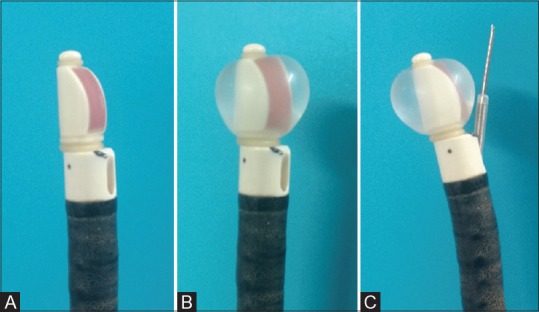
Intrathoracic lymphadenopathy is a common problem encountered in clinical practice and is caused by a wide variety of diseases. Traditionally, the mediastinal lymph nodes were sampled using conventional transbronchial needle aspiration (TBNA), or surgical methods such as mediastinoscopy, and thoracotomy (open or video-assisted thoracoscopy). However, surgical modalities including mediastinoscopy are invasive, expensive, and not universally available. Moreover, they are associated with considerable morbidity and mortality. Conventional TBNA although minimally invasive has a low diagnostic yield. In the last decade, endobronchial ultrasound-guided TBNA (EBUS-TBNA) has emerged as the diagnostic procedure of choice in evaluating undiagnosed intrathoracic lymphadenopathy. EBUS-TBNA is also currently the preferred modality in the mediastinal staging of lung cancer. The procedure is minimally invasive, safe, and can be performed as a day-care procedure. In the era of personalized medicine in lung cancer, optimizing the procedure, sample collection, and processing are crucial, as more tissue is required for performing a wide array of molecular tests. Despite its widespread use and acceptance, the diagnostic sensitivity of EBUS-TBNA is still low. To maximize the yield, cytologists and physicians should be aware of the technical details of the procedure. Herein, we discuss the technique of performing EBUS-TBNA, its indications, contraindications, and the processing of the samples at our bronchoscopy suite. We also highlight the challenges faced by the cytologists and clinicians while processing EBUS aspirates.
胸内淋巴结病是临床实践中常见的问题,由多种疾病引起。传统上,纵隔淋巴结采样采用传统的经支气管针吸活检(TBNA),或采用诸如纵隔镜检查和开胸手术(开放或电视辅助胸腔镜手术)等手术方法。然而,包括纵隔镜检查在内的手术方式具有侵入性、费用高且并非普遍可用。此外,它们还伴有相当高的发病率和死亡率。传统的TBNA虽然微创,但诊断率较低。在过去十年中,支气管内超声引导下经支气管针吸活检(EBUS-TBNA)已成为评估未确诊的胸内淋巴结病的首选诊断方法。EBUS-TBNA目前也是肺癌纵隔分期的首选方式。该操作微创、安全,可作为日间手术进行。在肺癌个体化医疗时代,优化操作、样本采集和处理至关重要,因为进行一系列分子检测需要更多组织。尽管EBUS-TBNA已被广泛应用和接受,但其诊断敏感性仍然较低。为了最大限度地提高诊断率,细胞学家和医生应了解该操作的技术细节。在此,我们讨论在我们的支气管镜检查室进行EBUS-TBNA的技术、其适应证、禁忌证以及样本处理。我们还强调了细胞学家和临床医生在处理EBUS吸出物时面临的挑战。