The First Clinical Medical School of Lanzhou University, Lanzhou, Gansu 730000, P.R. China.
Department of Special Minimally Invasive Surgery, The First Hospital of Lanzhou University, Lanzhou, Gansu 730000, P.R. China.
Oncol Rep. 2019 Mar;41(3):1539-1548. doi: 10.3892/or.2019.6977. Epub 2019 Jan 22.
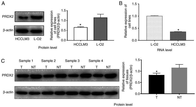
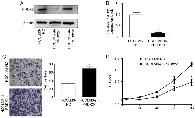
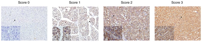
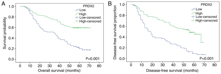
It has been revealed by our previous proteomic study that the expression profile is different between well‑differentiated and poorly differentiated hepatocellular carcinoma (HCC). Among those differently expressed proteins, peroxiredoxin2 (PRDX2) was our protein of interest. The present study aimed to further investigate the value of PRDX2 as a prognostic factor in HCC. Tissue microarrays were used to investigate the expression difference between HCC tissues and their adjacent normal liver tissues. The expression of PRDX2 at both mRNA and protein levels was examined by q‑RT‑PCR, western blotting and immunohistochemical assessment in HCC tissues and cell line HCCLM3. Silencing of PRDX2 in HCCLM3 was achieved usingpGMLV‑SC1 lentiviral vectors. Cell Counting Kit‑8 (CCK‑8) and Transwell migration assays were used to assess cell proliferation and migration, respectively. Categorical variables were assessed using the Chi‑square test, and ordinal variables were examined using the Mann‑Whitney U test. The difference of continuous variables between groups were compared with t‑tests. The Kaplan‑Meier method was used to calculate the overall survival (OS) and disease‑free survival (DFS) of patients, and the log‑rank test was used to analyze the differences between groups. The results revealed that the expression of PRDX2 was decreased at both the mRNA and protein levels in an HCC cell line compared to that of a normal human liver cell line. PRDX2 protein expression levels were significantly downregulated in HCC tissues and were positively linked to overall survival (OS) and disease‑free survival (DFS) of HCC patients. Patients with high PRDX2 expression levels had longer OS and DFS times than those with lower PRDX2 expression. Silencing of PRDX2 in the HCC cell line HCCLM3 promoted cancer cell proliferation and migration. Our findings indicated that PRDX2 may play an important role in HCC development; PRDX2 may serve as a useful prognostic factor and a therapeutic target.
我们之前的蛋白质组学研究表明,高分化和低分化肝细胞癌(HCC)之间的表达谱存在差异。在这些差异表达的蛋白质中,过氧化物还原酶 2(PRDX2)是我们感兴趣的蛋白质。本研究旨在进一步探讨 PRDX2 作为 HCC 预后因素的价值。组织微阵列用于研究 HCC 组织与其相邻正常肝组织之间的表达差异。通过 q-RT-PCR、western blot 和免疫组织化学评估,检测 HCC 组织和细胞系 HCCLM3 中 PRDX2 的表达。使用 pGMLV-SC1 慢病毒载体沉默 HCCLM3 中的 PRDX2。使用细胞计数试剂盒-8(CCK-8)和 Transwell 迁移实验分别评估细胞增殖和迁移。使用卡方检验评估分类变量,使用曼-惠特尼 U 检验评估有序变量。组间连续变量的差异用 t 检验进行比较。Kaplan-Meier 法用于计算患者的总生存(OS)和无病生存(DFS),对数秩检验用于分析组间差异。结果显示,与正常人类肝细胞系相比,PRDX2 在 HCC 细胞系中的 mRNA 和蛋白水平均降低。PRDX2 蛋白表达水平在 HCC 组织中显著下调,与 HCC 患者的总生存(OS)和无病生存(DFS)呈正相关。PRDX2 高表达患者的 OS 和 DFS 时间长于 PRDX2 低表达患者。沉默 HCC 细胞系 HCCLM3 中的 PRDX2 促进了癌细胞的增殖和迁移。我们的研究结果表明,PRDX2 可能在 HCC 发展中发挥重要作用;PRDX2 可能成为一种有用的预后因素和治疗靶点。