• 文献检索
  • 文档翻译
  • 深度研究
  • 学术资讯
  • Suppr Zotero 插件Zotero 插件
  • 邀请有礼
  • 套餐&价格
  • 历史记录
应用&插件
Suppr Zotero 插件Zotero 插件浏览器插件Mac 客户端Windows 客户端微信小程序
定价
高级版会员购买积分包购买API积分包
服务
文献检索文档翻译深度研究API 文档MCP 服务
关于我们
关于 Suppr公司介绍联系我们用户协议隐私条款
关注我们

Suppr 超能文献

核心技术专利:CN118964589B侵权必究
粤ICP备2023148730 号-1Suppr @ 2026

文献检索

告别复杂PubMed语法,用中文像聊天一样搜索,搜遍4000万医学文献。AI智能推荐,让科研检索更轻松。

立即免费搜索

文件翻译

保留排版,准确专业,支持PDF/Word/PPT等文件格式,支持 12+语言互译。

免费翻译文档

深度研究

AI帮你快速写综述,25分钟生成高质量综述,智能提取关键信息,辅助科研写作。

立即免费体验

一个生物物理模型解释了发育中的视网膜中的自发爆发行为。

A biophysical model explains the spontaneous bursting behavior in the developing retina.

机构信息

Biovision team, Université Côte d'Azur, Inria, France.

INPHYNI, Université Côte d'Azur, CNRS, Sophia Antipolis, France.

出版信息

Sci Rep. 2019 Feb 12;9(1):1859. doi: 10.1038/s41598-018-38299-4.

DOI:10.1038/s41598-018-38299-4
PMID:30755684
原文链接:https://pmc.ncbi.nlm.nih.gov/articles/PMC6372601/
Abstract

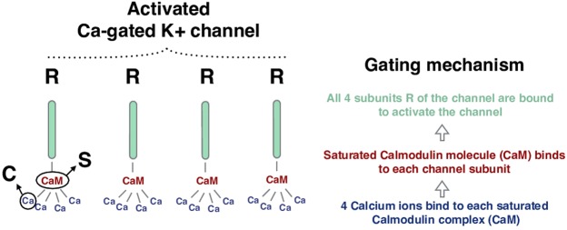
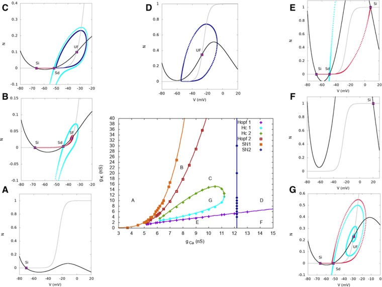
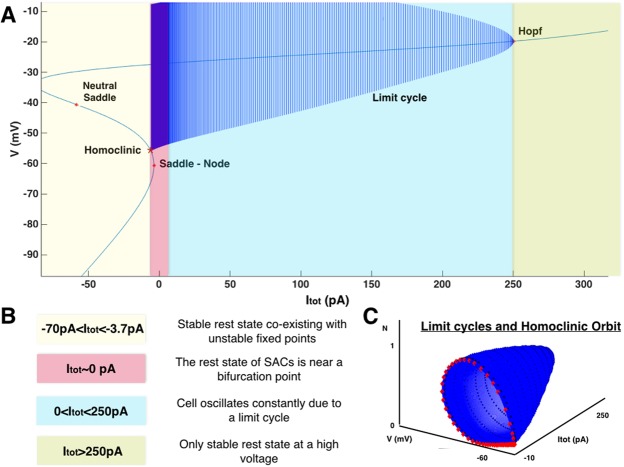
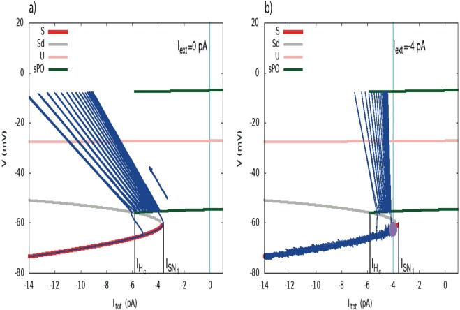
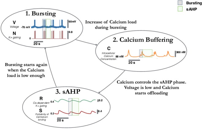
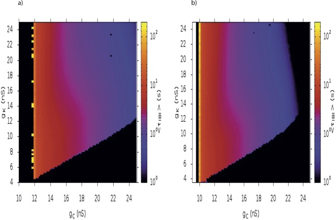
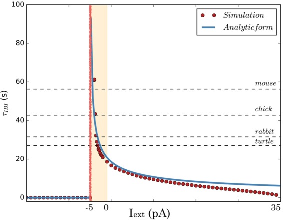
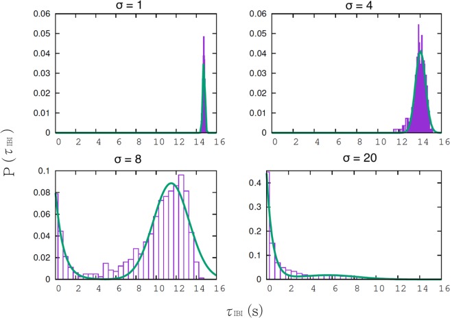
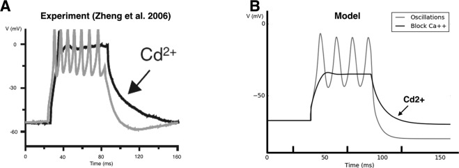
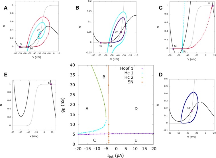
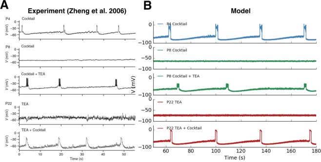
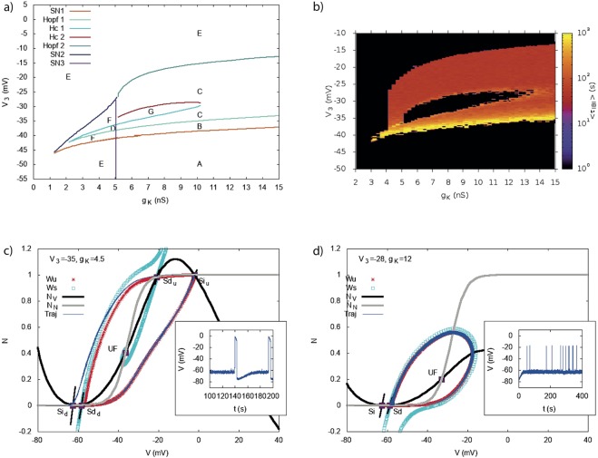
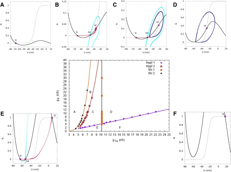
During early development, waves of activity propagate across the retina and play a key role in the proper wiring of the early visual system. During a particular phase of the retina development (stage II) these waves are triggered by a transient network of neurons, called Starburst Amacrine Cells (SACs), showing a bursting activity which disappears upon further maturation. The underlying mechanisms of the spontaneous bursting and the transient excitability of immature SACs are not completely clear yet. While several models have attempted to reproduce retinal waves, none of them is able to mimic the rhythmic autonomous bursting of individual SACs and reveal how these cells change their intrinsic properties during development. Here, we introduce a mathematical model, grounded on biophysics, which enables us to reproduce the bursting activity of SACs and to propose a plausible, generic and robust, mechanism that generates it. The core parameters controlling repetitive firing are fast depolarizing V-gated calcium channels and hyperpolarizing V-gated potassium channels. The quiescent phase of bursting is controlled by a slow after hyperpolarization (sAHP), mediated by calcium-dependent potassium channels. Based on a bifurcation analysis we show how biophysical parameters, regulating calcium and potassium activity, control the spontaneously occurring fast oscillatory activity followed by long refractory periods in individual SACs. We make a testable experimental prediction on the role of voltage-dependent potassium channels on the excitability properties of SACs and on the evolution of this excitability along development. We also propose an explanation on how SACs can exhibit a large variability in their bursting periods, as observed experimentally within a SACs network as well as across different species, yet based on a simple, unique, mechanism. As we discuss, these observations at the cellular level have a deep impact on the retinal waves description.

摘要

在早期发育过程中,活动波在视网膜中传播,并在早期视觉系统的正确布线中发挥关键作用。在视网膜发育的特定阶段(阶段 II),这些波是由称为星爆型无长突细胞(Starburst Amacrine Cells,SAC)的短暂神经元网络触发的,这些细胞表现出爆发活动,随着进一步成熟而消失。自发性爆发和不成熟 SACs 的短暂兴奋性的潜在机制尚不完全清楚。虽然已经有几个模型试图复制视网膜波,但没有一个能够模拟单个 SAC 的节律性自主爆发,并揭示这些细胞在发育过程中如何改变其内在特性。在这里,我们引入了一个基于生物物理学的数学模型,该模型使我们能够复制 SAC 的爆发活动,并提出一个合理、通用且稳健的机制来产生这种活动。控制重复放电的核心参数是快速去极化 V 门控钙通道和超极化 V 门控钾通道。爆发的静息期由钙依赖性钾通道介导的慢后超极化(slow after hyperpolarization,sAHP)控制。基于分岔分析,我们展示了调节钙和钾活性的生物物理参数如何控制单个 SACs 中自发发生的快速振荡活动,随后是长时间的不应期。我们对电压依赖性钾通道在 SACs 兴奋性特性上的作用以及这种兴奋性在发育过程中的演变提出了可测试的实验预测。我们还提出了一种解释,说明 SACs 如何表现出其爆发周期的大变化,正如在 SACs 网络以及不同物种中观察到的那样,但基于一种简单、独特的机制。正如我们所讨论的,这些细胞水平的观察对视网膜波的描述有深远的影响。

https://cdn.ncbi.nlm.nih.gov/pmc/blobs/59ed/6372601/bba766f70b3a/41598_2018_38299_Fig13_HTML.jpg
https://cdn.ncbi.nlm.nih.gov/pmc/blobs/59ed/6372601/8e23611c9b89/41598_2018_38299_Fig1_HTML.jpg
https://cdn.ncbi.nlm.nih.gov/pmc/blobs/59ed/6372601/574f74682c3b/41598_2018_38299_Fig2_HTML.jpg
https://cdn.ncbi.nlm.nih.gov/pmc/blobs/59ed/6372601/f88fc68e8606/41598_2018_38299_Fig3_HTML.jpg
https://cdn.ncbi.nlm.nih.gov/pmc/blobs/59ed/6372601/709eb5ca26f7/41598_2018_38299_Fig4_HTML.jpg
https://cdn.ncbi.nlm.nih.gov/pmc/blobs/59ed/6372601/3f564b0383c9/41598_2018_38299_Fig5_HTML.jpg
https://cdn.ncbi.nlm.nih.gov/pmc/blobs/59ed/6372601/9440e8ad57a1/41598_2018_38299_Fig6_HTML.jpg
https://cdn.ncbi.nlm.nih.gov/pmc/blobs/59ed/6372601/873d41b85728/41598_2018_38299_Fig7_HTML.jpg
https://cdn.ncbi.nlm.nih.gov/pmc/blobs/59ed/6372601/f8a73561e46d/41598_2018_38299_Fig8_HTML.jpg
https://cdn.ncbi.nlm.nih.gov/pmc/blobs/59ed/6372601/74a8bf81255b/41598_2018_38299_Fig9_HTML.jpg
https://cdn.ncbi.nlm.nih.gov/pmc/blobs/59ed/6372601/6c4f67f1fd50/41598_2018_38299_Fig10_HTML.jpg
https://cdn.ncbi.nlm.nih.gov/pmc/blobs/59ed/6372601/39656905a179/41598_2018_38299_Fig11_HTML.jpg
https://cdn.ncbi.nlm.nih.gov/pmc/blobs/59ed/6372601/2e1855c40f97/41598_2018_38299_Fig12_HTML.jpg
https://cdn.ncbi.nlm.nih.gov/pmc/blobs/59ed/6372601/bba766f70b3a/41598_2018_38299_Fig13_HTML.jpg

相似文献

1
A biophysical model explains the spontaneous bursting behavior in the developing retina.一个生物物理模型解释了发育中的视网膜中的自发爆发行为。
Sci Rep. 2019 Feb 12;9(1):1859. doi: 10.1038/s41598-018-38299-4.
2
Retinal Wave Patterns Are Governed by Mutual Excitation among Starburst Amacrine Cells and Drive the Refinement and Maintenance of Visual Circuits.视网膜波模式受无长突星形细胞之间的相互兴奋作用支配,并驱动视觉回路的精细化和维持。
J Neurosci. 2016 Mar 30;36(13):3871-86. doi: 10.1523/JNEUROSCI.3549-15.2016.
3
A transient network of intrinsically bursting starburst cells underlies the generation of retinal waves.由具有内在爆发性的星爆细胞组成的瞬态网络是视网膜波产生的基础。
Nat Neurosci. 2006 Mar;9(3):363-71. doi: 10.1038/nn1644. Epub 2006 Feb 5.
4
Cellular mechanisms underlying spatiotemporal features of cholinergic retinal waves.胆碱能视网膜波时空特征的细胞机制。
J Neurosci. 2012 Jan 18;32(3):850-63. doi: 10.1523/JNEUROSCI.5309-12.2012.
5
Presynaptic SNAP-25 regulates retinal waves and retinogeniculate projection via phosphorylation.突触前 SNAP-25 通过磷酸化调节视网膜波和视网膜-外侧膝状体投射。
Proc Natl Acad Sci U S A. 2019 Feb 19;116(8):3262-3267. doi: 10.1073/pnas.1812169116. Epub 2019 Feb 6.
6
Spontaneous Depolarization-Induced Action Potentials of ON-Starburst Amacrine Cells during Cholinergic and Glutamatergic Retinal Waves.胆碱能和谷氨酸能视网膜波期间ON-星爆无长突细胞的自发去极化诱导动作电位
Cells. 2020 Dec 1;9(12):2574. doi: 10.3390/cells9122574.
7
Adenosine A(2A) receptor up-regulates retinal wave frequency via starburst amacrine cells in the developing rat retina.在发育中的大鼠视网膜中,腺苷A(2A)受体通过无长突星形细胞上调视网膜波频率。
PLoS One. 2014 Apr 28;9(4):e95090. doi: 10.1371/journal.pone.0095090. eCollection 2014.
8
A unique role for Kv3 voltage-gated potassium channels in starburst amacrine cell signaling in mouse retina.Kv3电压门控钾通道在小鼠视网膜星爆无长突细胞信号传导中的独特作用。
J Neurosci. 2004 Aug 18;24(33):7335-43. doi: 10.1523/JNEUROSCI.1275-04.2004.
9
A role for TREK1 in generating the slow afterhyperpolarization in developing starburst amacrine cells.TREK1 在产生暴发性星形胶质细胞慢后超极化中的作用。
J Neurophysiol. 2013 May;109(9):2250-9. doi: 10.1152/jn.01085.2012. Epub 2013 Feb 6.
10
Elucidating the role of AII amacrine cells in glutamatergic retinal waves.阐明无长突细胞AII在视网膜谷氨酸能波中的作用。
J Neurosci. 2015 Jan 28;35(4):1675-86. doi: 10.1523/JNEUROSCI.3291-14.2015.

引用本文的文献

1
Modeling cholinergic retinal waves: starburst amacrine cells shape wave generation, propagation, and direction bias.建模胆碱能视网膜波:星形胶质细胞调节波的产生、传播和方向偏向。
Sci Rep. 2023 Feb 17;13(1):2834. doi: 10.1038/s41598-023-29572-2.
2
Retinal Processing: Insights from Mathematical Modelling.视网膜处理:数学建模的见解
J Imaging. 2022 Jan 17;8(1):14. doi: 10.3390/jimaging8010014.
3
On the potential role of lateral connectivity in retinal anticipation.关于视网膜预期中侧向连接的潜在作用。

本文引用的文献

1
A mathematical model of calcium dynamics in HSY cells.HSY细胞中钙动力学的数学模型。
PLoS Comput Biol. 2017 Feb 15;13(2):e1005275. doi: 10.1371/journal.pcbi.1005275. eCollection 2017 Feb.
2
Retinal Wave Patterns Are Governed by Mutual Excitation among Starburst Amacrine Cells and Drive the Refinement and Maintenance of Visual Circuits.视网膜波模式受无长突星形细胞之间的相互兴奋作用支配,并驱动视觉回路的精细化和维持。
J Neurosci. 2016 Mar 30;36(13):3871-86. doi: 10.1523/JNEUROSCI.3549-15.2016.
3
A reaction-diffusion model of cholinergic retinal waves.
J Math Neurosci. 2021 Jan 9;11(1):3. doi: 10.1186/s13408-020-00101-z.
4
Theoretical Models of Neural Development.神经发育的理论模型
iScience. 2018 Oct 26;8:183-199. doi: 10.1016/j.isci.2018.09.017. Epub 2018 Sep 27.
胆碱能视网膜波的反应扩散模型。
PLoS Comput Biol. 2014 Dec 4;10(12):e1003953. doi: 10.1371/journal.pcbi.1003953. eCollection 2014 Dec.
4
Intrinsic bursting of AII amacrine cells underlies oscillations in the rd1 mouse retina.AII无长突细胞的内在爆发是rd1小鼠视网膜振荡的基础。
J Neurophysiol. 2014 Sep 15;112(6):1491-504. doi: 10.1152/jn.00437.2014. Epub 2014 Jul 9.
5
Following the ontogeny of retinal waves: pan-retinal recordings of population dynamics in the neonatal mouse.视网膜波的发生发展:新生期小鼠视网膜群体动力学的全视网膜记录。
J Physiol. 2014 Apr 1;592(7):1545-63. doi: 10.1113/jphysiol.2013.262840. Epub 2013 Dec 23.
6
A theory of plasma membrane calcium pump stimulation and activity.质膜钙泵刺激与活性的理论
J Biol Phys. 2005 May;31(2):183-206. doi: 10.1007/s10867-005-4472-2.
7
Cooperation of intrinsic bursting and calcium oscillations underlying activity patterns of model pre-Bötzinger complex neurons.前包钦格复合体模型神经元活动模式背后的内在爆发与钙振荡的协同作用。
J Comput Neurosci. 2013 Apr;34(2):345-66. doi: 10.1007/s10827-012-0425-5. Epub 2012 Sep 28.
8
Hybrid models and biological model reduction with PyDSTool.基于 PyDSTool 的混合模型与生物模型降阶。
PLoS Comput Biol. 2012;8(8):e1002628. doi: 10.1371/journal.pcbi.1002628. Epub 2012 Aug 9.
9
Cellular mechanisms underlying spatiotemporal features of cholinergic retinal waves.胆碱能视网膜波时空特征的细胞机制。
J Neurosci. 2012 Jan 18;32(3):850-63. doi: 10.1523/JNEUROSCI.5309-12.2012.
10
Assembly and disassembly of a retinal cholinergic network.视网膜胆碱能网络的组装与拆卸。
Vis Neurosci. 2012 Jan;29(1):61-71. doi: 10.1017/S0952523811000216. Epub 2011 Jul 26.