Laboratory of Pathology, National Cancer Institute, National Institute of Health, Bethesda, MD, USA.
Department of Molecular Diagnostics, Holycross Cancer Center, Kielce, Poland.
Mod Pathol. 2019 Jul;32(7):957-966. doi: 10.1038/s41379-018-0163-y. Epub 2019 Feb 13.
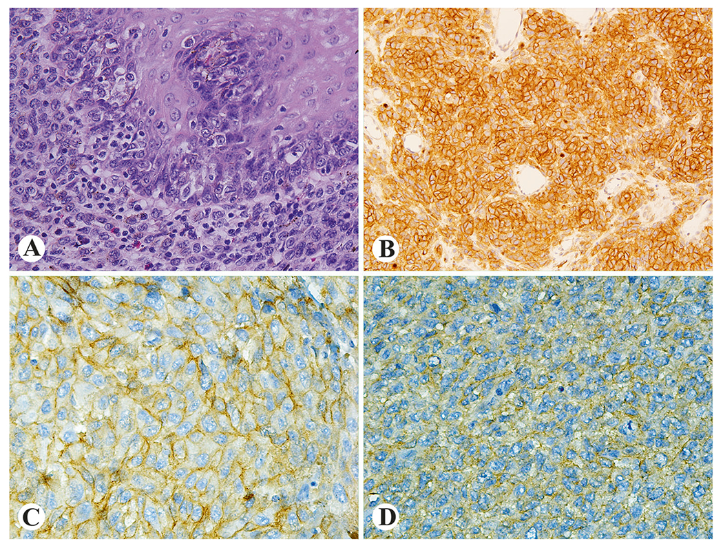
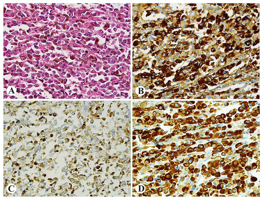
Primary malignant melanoma of esophagus is very rare, and its clinicopathologic and genetic features have not been extensively investigated. In this study, 20 tumors from 14 male and 6 female patients (40-79 years old) were evaluated. Dysphagia, chest pain, and weight loss were frequent symptoms. Thirteen melanomas, including two with multiple lesions, involved the distal third of esophagus. The median tumor diameter was 6 cm. Epithelioid morphology, moderate atypia, and pigmentation were typical findings. None of the patients had melanoma elsewhere, and all tumors exhibited a junctional peri-epithelial component consistent with a primary lesion. The median mitotic activity was 11 per 10 high-power fields (range, 0-31). Nine patients died of tumor within 4-22 months, however, two showed long-term (96 and 104 months) survival. In 15 cases, tissue for further immunohistochemical and molecular studies were available. BRAF, KIT, and NRAS mutation status was assessed by Sanger sequencing in all 15 tumors. The next-generation sequencing of 50 or 409 genes was performed in five and three cases, respectively. IGF1R expression indicating activation of the IGF axis was seen in 82% (9/11) of tumors. However, no BRAF mutations were identified. In 33% (5/15) of tumors, NRAS mutations were detected. KIT expression was seen in 50% (7/14) of melanomas including single KIT mutant. Two of three tumors evaluated with 409 genes panel revealed multiple driver mutations indicating sub-clonal expansion, whereas a single mutation (TSC1 p.H371Q) was the sole change in the third case. SF3B1 p.K666T and p.R625C mutations were detected in two cases. However, no co-occurrence of SF3B1 and GNAQ or GNA11 mutations, seen in uveal melanoma, was detected. FBXW7 p.R465C and p.R479G mutations, linked to cancer progression, were found in two of eight tumors. In summary, esophageal melanoma mutation profile indicates complexity of molecular mechanisms underlying its pathogenesis.
原发性食管恶性黑色素瘤非常罕见,其临床病理和遗传特征尚未得到广泛研究。在这项研究中,评估了来自 14 名男性和 6 名女性患者(40-79 岁)的 20 个肿瘤。吞咽困难、胸痛和体重减轻是常见的症状。13 个黑色素瘤,包括 2 个多发性病变,累及食管远端三分之一。肿瘤的中位直径为 6cm。上皮样形态、中度异型性和色素沉着是典型的发现。没有患者有其他部位的黑色素瘤,所有肿瘤均表现出与原发性病变一致的上皮下交界性成分。中位有丝分裂活性为每 10 个高倍视野 11 个(范围 0-31)。9 例患者在 4-22 个月内因肿瘤死亡,但 2 例患者长期(96 和 104 个月)存活。在 15 例病例中,有进一步进行免疫组织化学和分子研究的组织。对所有 15 个肿瘤进行 Sanger 测序评估 BRAF、KIT 和 NRAS 突变状态。对 5 个和 3 个病例分别进行了 50 个或 409 个基因的下一代测序。在 82%(9/11)的肿瘤中观察到 IGF1R 表达,表明 IGF 轴的激活。然而,没有发现 BRAF 突变。在 33%(5/15)的肿瘤中,检测到 NRAS 突变。KIT 表达见于 50%(14/28)的黑色素瘤,包括单个 KIT 突变。在 3 个用 409 个基因面板评估的肿瘤中,有 2 个肿瘤显示出多个驱动突变,表明亚克隆扩张,而第 3 个肿瘤只有一个突变(TSC1 p.H371Q)。在 2 个病例中检测到 SF3B1 p.K666T 和 p.R625C 突变。然而,在葡萄膜黑色素瘤中发现的 SF3B1 和 GNAQ 或 GNA11 突变的共同发生并未在本研究中发现。FBXW7 p.R465C 和 p.R479G 突变与癌症进展有关,在 8 个肿瘤中的 2 个中发现。总之,食管黑色素瘤的突变谱表明其发病机制的分子机制的复杂性。