Privolzhsky Research Medical University, 603950 Minin and Pozharsky Sq., 10/1, Nizhny Novgorod, Russia.
Institute of Applied Physics, Russian Academy of Sciences, 603950 Ulyanova Str., 46, Nizhny Novgorod, Russia.
Sci Rep. 2019 Feb 14;9(1):2024. doi: 10.1038/s41598-019-38493-y.
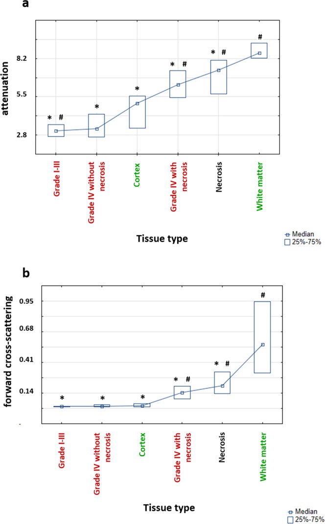
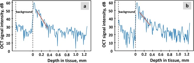
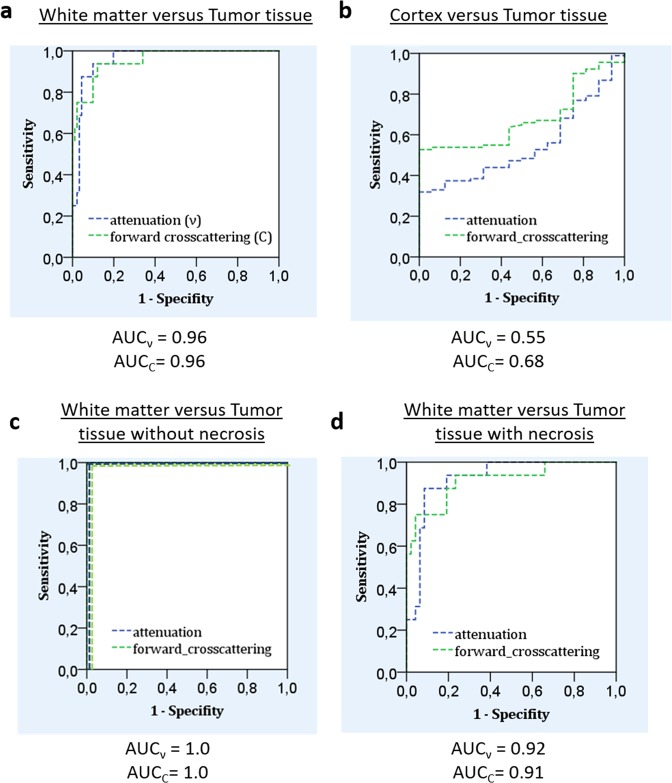
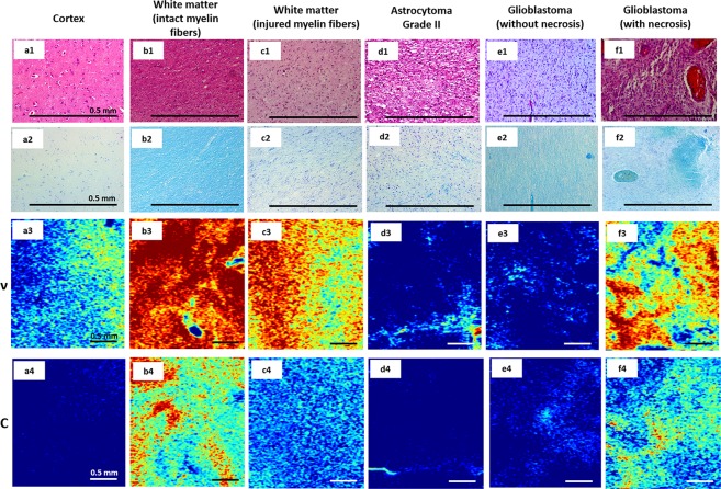
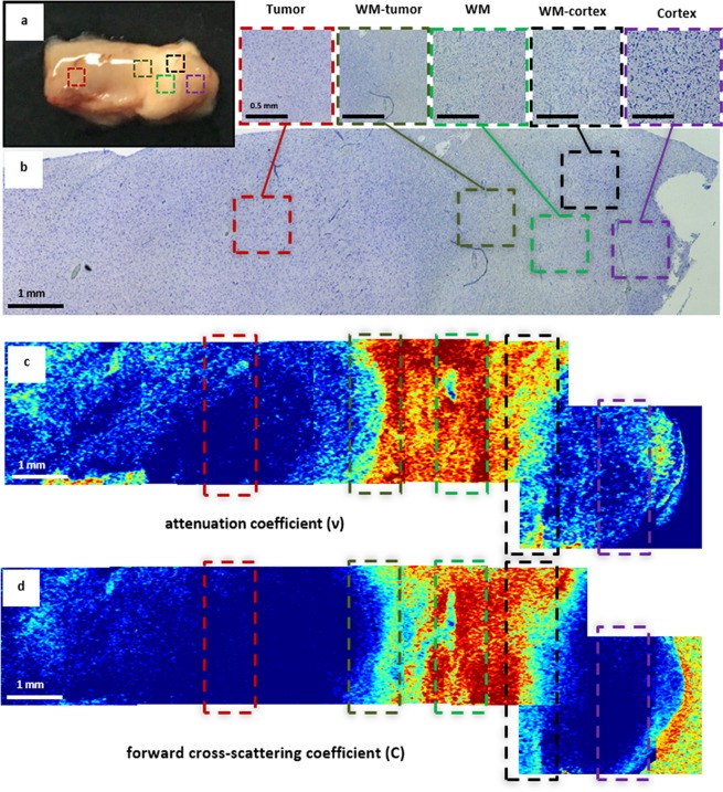
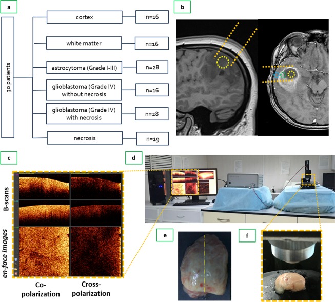
Optical coherence tomography (OCT) is a promising method for detecting cancer margins during tumor resection. This study focused on differentiating tumorous from nontumorous tissues in human brain tissues using cross-polarization OCT (CP OCT). The study was performed on fresh ex vivo human brain tissues from 30 patients with high- and low-grade gliomas. Different tissue types that neurosurgeons should clearly distinguish during surgery, such as the cortex, white matter, necrosis and tumorous tissue, were separately analyzed. Based on volumetric CP OCT data, tumorous and normal brain tissue were differentiated using two optical coefficients - attenuation and forward cross-scattering. Compared with white matter, tumorous tissue without necrotic areas had significantly lower optical attenuation and forward cross-scattering values. The presence of particular morphological patterns, such as necrosis and injured myelinated fibers, can lead to dramatic changes in coefficient values and create some difficulties in differentiating between tissues. Color-coded CP OCT maps based on optical coefficients provided a visual assessment of the tissue. This study demonstrated the high translational potential of CP OCT in differentiating tumorous tissue from white matter. The clinical use of CP OCT during surgery in patients with gliomas could increase the extent of tumor resection and improve overall and progression-free survival.
光学相干断层扫描(OCT)是一种很有前途的方法,可用于在肿瘤切除过程中检测肿瘤边缘。本研究专注于使用交叉偏振 OCT(CP OCT)来区分人脑组织中的肿瘤组织和非肿瘤组织。该研究在 30 名高级和低级脑胶质瘤患者的新鲜离体人脑组织上进行。对神经外科医生在手术中需要清楚区分的不同组织类型,如皮质、白质、坏死组织和肿瘤组织,分别进行了分析。基于体积 CP OCT 数据,使用两个光学系数 - 衰减和前向交叉散射来区分肿瘤组织和正常脑组织。与白质相比,无坏死区域的肿瘤组织的光衰减和前向交叉散射值显著降低。特定形态模式(如坏死和受损的髓鞘纤维)的存在会导致系数值发生剧烈变化,并在组织区分方面造成一些困难。基于光学系数的彩色 CP OCT 图谱提供了组织的直观评估。本研究证明了 CP OCT 在区分肿瘤组织和白质方面具有很高的转化潜力。CP OCT 在脑胶质瘤患者手术中的临床应用可以增加肿瘤切除范围,并提高整体和无进展生存率。