Hubrecht Institute-KNAW & University Medical Center Utrecht, Uppsalalaan 8, 3584 CT, Utrecht, The Netherlands.
Prinses Máxima Center for Pediatric Oncology, Uppsalalaan 8, 3584CT, Utrecht, The Netherlands.
Sci Rep. 2019 Feb 14;9(1):2054. doi: 10.1038/s41598-019-38625-4.
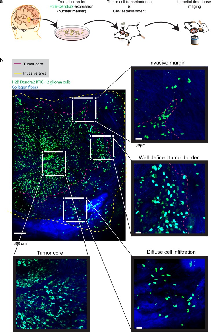
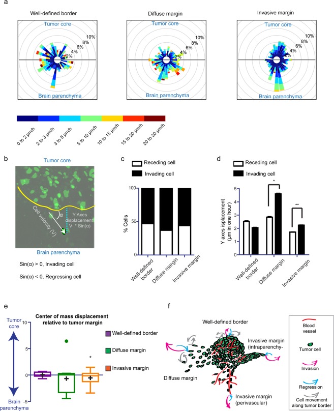
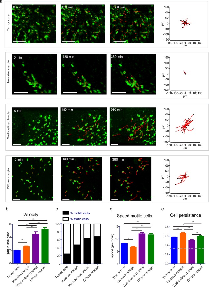
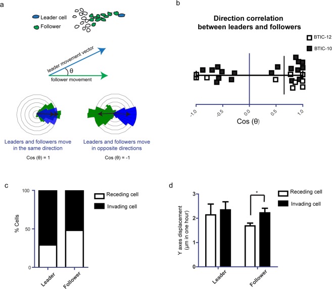
The pathogenesis of glioblastoma (GBM) is characterized by highly invasive behavior allowing dissemination and progression. A conclusive image of the invasive process is not available. The aim of this work was to study invasion dynamics in GBM using an innovative in vivo imaging approach. Primary brain tumor initiating cell lines from IDH-wild type GBM stably expressing H2B-Dendra2 were implanted orthotopically in the brains of SCID mice. Using high-resolution time-lapse intravital imaging, tumor cell migration in the tumor core, border and invasive front was recorded. Tumor cell dynamics at different border configurations were analyzed and multivariate linear modelling of tumor cell spreading was performed. We found tumor border configurations, recapitulating human tumor border morphologies. Not only tumor borders but also the tumor core was composed of highly dynamic cells, with no clear correlation to the ability to spread into the brain. Two types of border configurations contributed to tumor cell spreading through distinct invasion patterns: an invasive margin that executes slow but directed invasion, and a diffuse infiltration margin with fast but less directed movement. By providing a more detailed view on glioma invasion patterns, our study may improve accuracy of prognosis and serve as a basis for personalized therapeutic approaches.
胶质母细胞瘤(GBM)的发病机制以高度侵袭性行为为特征,允许其扩散和进展。目前还没有明确的侵袭过程图像。本研究旨在使用创新的体内成像方法研究 GBM 的侵袭动力学。从 IDH-野生型 GBM 中分离出的原代脑肿瘤起始细胞系,稳定表达 H2B-Dendra2,然后原位植入 SCID 小鼠的大脑中。使用高分辨率延时活体成像,记录肿瘤核心、边界和侵袭前沿的肿瘤细胞迁移。分析了不同边界构型下的肿瘤细胞动力学,并对肿瘤细胞扩散进行了多元线性建模。我们发现了肿瘤边界构型,再现了人类肿瘤边界形态。不仅肿瘤边界,而且肿瘤核心都是由高度动态的细胞组成的,与向大脑扩散的能力没有明显的相关性。两种边界构型通过不同的侵袭模式促进肿瘤细胞的扩散:具有缓慢但定向侵袭能力的侵袭边界,以及具有快速但定向运动能力较弱的弥散浸润边界。通过更详细地观察神经胶质瘤的侵袭模式,本研究可能会提高预后的准确性,并为个性化治疗方法提供依据。