Harlalka Vatika, Bapi Raju S, Vinod P K, Roy Dipanjan
Center for Computational Natural Sciences and Bioinformatics, IIIT Hyderabad, Hyderabad, India.
Cognitive Science Lab, IIIT Hyderabad, Hyderabad, India.
Front Hum Neurosci. 2019 Feb 1;13:6. doi: 10.3389/fnhum.2019.00006. eCollection 2019.
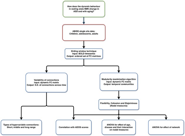
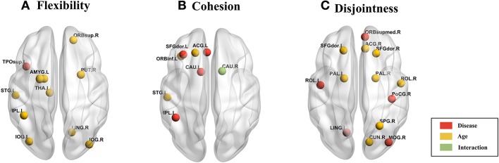
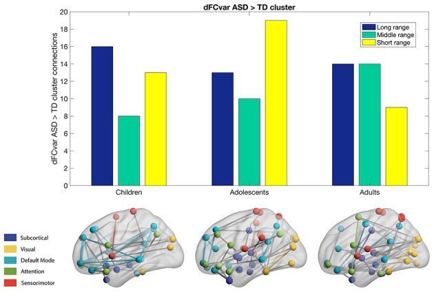
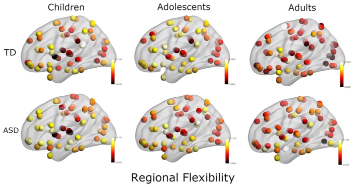
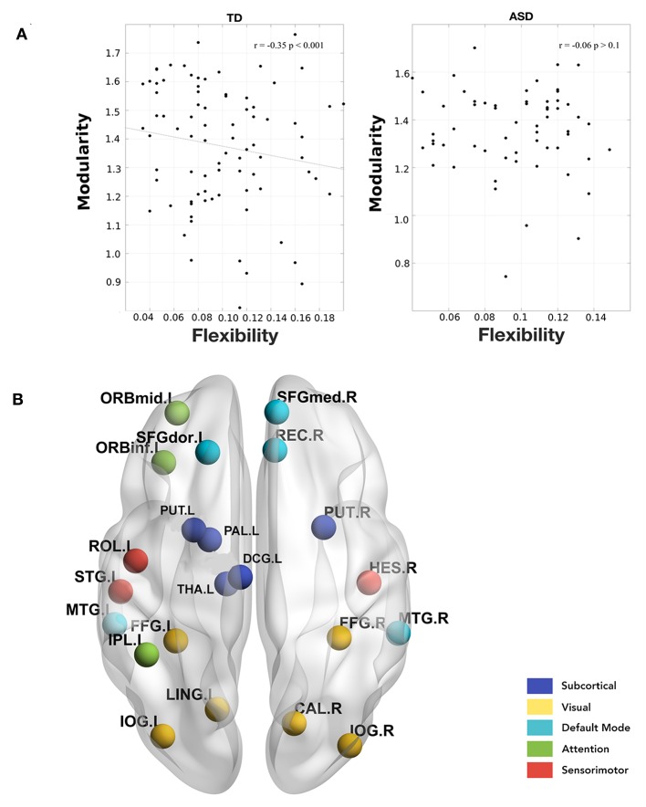
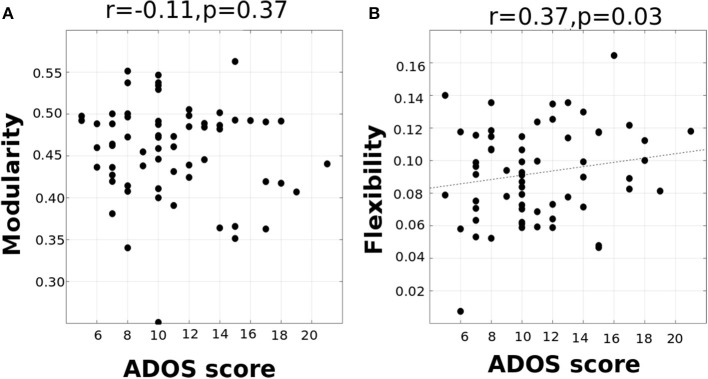
Resting-state functional connectivity (FC) analyses have shown atypical connectivity in autism spectrum disorder (ASD) as compared to typically developing (TD). However, this view emerges from investigating static FC overlooking the whole brain transient connectivity patterns. In our study, we investigated how age and disease influence the dynamic changes in functional connectivity of TD and ASD. We used resting-state functional magnetic resonance imaging (rs-fMRI) data stratified into three cohorts: children (7-11 years), adolescents (12-17 years), and adults (18+ years) for the analysis. The dynamic variability in the connection strength and the modular organization in terms of measures such as flexiblity, cohesion strength, and disjointness were explored for each subject to characterize the differences between ASD and TD. In ASD, we observed significantly higher inter-subject dynamic variability in connection strength as compared to TD. This hyper-variability relates to the symptom severity in ASD. We also found that whole-brain flexibility correlates with static modularity only in TD. Further, we observed a core-periphery organization in the resting-state, with Sensorimotor and Visual regions in the rigid core; and DMN and attention areas in the flexible periphery. TD also develops a more cohesive organization of sensorimotor areas. However, in ASD we found a strong positive correlation of symptom severity with flexibility of rigid areas and with disjointness of sensorimotor areas. The regions of the brain showing high predictive power of symptom severity were distributed across the cortex, with stronger bearings in the frontal, motor, and occipital cortices. Our study demonstrates that the dynamic framework best characterizes the variability in ASD.
静息态功能连接(FC)分析表明,与典型发育(TD)人群相比,自闭症谱系障碍(ASD)患者存在非典型连接。然而,这一观点源于对静态FC的研究,忽视了全脑的瞬态连接模式。在我们的研究中,我们调查了年龄和疾病如何影响TD和ASD患者功能连接的动态变化。我们使用静息态功能磁共振成像(rs-fMRI)数据,将其分为三个队列:儿童(7 - 11岁)、青少年(12 - 17岁)和成人(18岁以上)进行分析。针对每个受试者,探索连接强度的动态变异性以及诸如灵活性、凝聚强度和不相交性等指标所衡量的模块化组织,以表征ASD和TD之间的差异。在ASD患者中,我们观察到与TD相比,受试者间连接强度的动态变异性显著更高。这种高变异性与ASD的症状严重程度相关。我们还发现,全脑灵活性仅在TD中与静态模块化相关。此外,我们在静息态中观察到一种核心 - 外围组织,感觉运动和视觉区域位于刚性核心;而默认模式网络(DMN)和注意力区域位于灵活外围。TD患者的感觉运动区域也发展出更具凝聚力的组织。然而,在ASD患者中,我们发现症状严重程度与刚性区域的灵活性以及感觉运动区域的不相交性呈强正相关。大脑中显示出对症状严重程度具有高预测力的区域分布在整个皮层,在额叶、运动皮层和枕叶皮层中影响更强。我们的研究表明,动态框架最能表征ASD中的变异性。