Institute of Neuro- and Sensory Physiology, University Medical Center Göttingen, Humboldtallee 23, 37073, Göttingen, Germany.
Center for Biostructural Imaging of Neurodegeneration (BIN), University of Göttingen Medical Center, von-Siebold-Straße 3a, 37075, Göttingen, Germany.
Nat Commun. 2019 Feb 18;10(1):820. doi: 10.1038/s41467-019-08677-1.
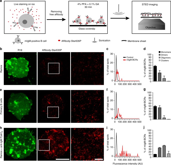
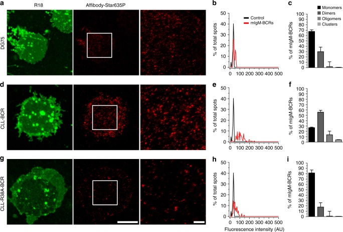
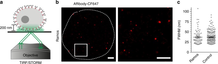
Stimulation of the B cell antigen receptor (BCR) triggers signaling pathways that promote the differentiation of B cells into plasma cells. Despite the pivotal function of BCR in B cell activation, the organization of the BCR on the surface of resting and antigen-activated B cells remains unclear. Here we show, using STED super-resolution microscopy, that IgM-containing BCRs exist predominantly as monomers and dimers in the plasma membrane of resting B cells, but form higher oligomeric clusters upon stimulation. By contrast, a chronic lymphocytic leukemia-derived BCR forms dimers and oligomers in the absence of a stimulus, but a single amino acid exchange reverts its organization to monomers in unstimulated B cells. Our super-resolution microscopy approach for quantitatively analyzing cell surface proteins may thus help reveal the nanoscale organization of immunoreceptors in various cell types.
刺激 B 细胞抗原受体 (BCR) 会触发信号通路,促进 B 细胞分化为浆细胞。尽管 BCR 在 B 细胞激活中起着关键作用,但静止和抗原激活的 B 细胞表面 BCR 的组织形式仍不清楚。在这里,我们使用 STED 超分辨率显微镜显示,在静止 B 细胞的质膜中,IgM 包含的 BCR 主要以单体和二聚体形式存在,但在受到刺激后会形成更高的寡聚体簇。相比之下,源自慢性淋巴细胞白血病的 BCR 在没有刺激的情况下形成二聚体和寡聚体,但单个氨基酸交换会使其在未受刺激的 B 细胞中恢复为单体。因此,我们用于定量分析细胞表面蛋白的超分辨率显微镜方法可能有助于揭示各种细胞类型中免疫受体的纳米级组织。