Gastroenterology Center, Ehime Prefectural Central Hospital, 83 Kasuga-cho, Matsuyama, Japan.
Department of Gastroenterology and Metabology, Ehime University Graduate School of Medicine, Toon, Japan.
J Cachexia Sarcopenia Muscle. 2019 Apr;10(2):347-354. doi: 10.1002/jcsm.12392. Epub 2019 Feb 21.
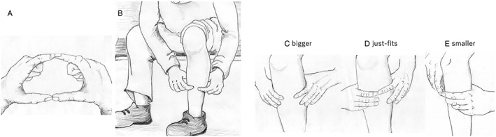
Muscle atrophy (MA) and muscle strength decline are important clinical features in chronic liver disease (CLD) patients. An easy to perform MA screening method without need for special equipment would be helpful. We evaluated the usefulness of the previously reported finger-circle test as screening for MA in CLD patients.
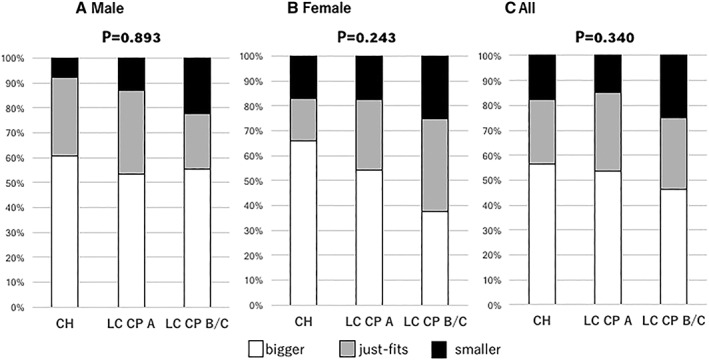
We retrospectively enrolled 358 Japanese CLD outpatients (70.8 ± 10.2 years, male/female = 234/124) who had undergone a computed tomography examination from December 2017 to March 2018, of whom 137 had chronic hepatitis, 169 had liver cirrhosis Child-Pugh A, and 52 had liver cirrhosis Child-Pugh B/C. Bilateral psoas muscle area at the middle of the third lumber vertebra (L3) was evaluated with computed tomography findings, which was performed as a screening of hepatocellular carcinoma, using a previously reported parameter for MA [psoas index (PI): total psoas muscle area (cm )/height (m) ] [mean PI ± standard deviation (SD) of male patients: 6.50 ± 1.13 cm /m and those of female patients: 4.30 ± 0.90 cm /m ]. We then evaluated the correlation between MA and finger-circle test results in these patients.
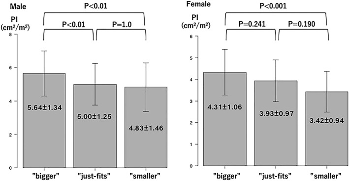
The mean PI values for finger-circle test results Bigger, Just-fits, and Smaller were 5.64 ± 1.34, 5.00 ± 1.25, and 4.83 ± 1.46 cm /m , respectively, in male patients (P < 0.001) and 4.31 ± 1.06, 3.93 ± 0.97, and 3.42 ± 0.94 cm /m , respectively, in female patients (P = 0.001). We found that a finger-circle test result in male patients other than Bigger (Just-fits and Smaller) predicted a decline in psoas muscle area of L3 to PI 5.25 cm /m (sensitivity/specificity 0.619/0.667, area under the curve 0.654, 95% confidence interval 0.583-0.724), which was approximately mean minus 1 SD (5.37 cm /m ). On the other hand, a Smaller test result in female patients predicted a decline in psoas muscle area of L3 to PI 3.33 cm /m (sensitivity/specificity 0.740/0.583, area under the curve 0.698, 95% confidence interval 0.583-0.813), approximately mean minus 1 SD (3.40 cm /m ).
The finger-circle test is an easy to perform and effective screening method for predicting earlier stage of MA in CLD patients without the need for special equipment.
肌肉萎缩(MA)和肌肉力量下降是慢性肝病(CLD)患者的重要临床特征。一种无需特殊设备即可进行的易于操作的 MA 筛查方法将很有帮助。我们评估了之前报道的手指圈试验作为 CLD 患者 MA 筛查的有用性。
我们回顾性纳入了 2017 年 12 月至 2018 年 3 月期间接受过计算机断层扫描检查的 358 例日本 CLD 门诊患者(70.8±10.2 岁,男性/女性=234/124),其中 137 例患有慢性肝炎,169 例患有 Child-Pugh A 级肝硬化,52 例患有 Child-Pugh B/C 级肝硬化。使用先前报道的 MA 指标[腰大肌指数(PI):总腰大肌面积(cm)/身高(m)]评估第 3 腰椎(L3)中部的双侧腰大肌面积,计算机断层扫描检查是作为肝细胞癌的筛查方法进行的[男性患者的平均 PI±标准差(SD):6.50±1.13 cm/m,女性患者的平均 PI±SD:4.30±0.90 cm/m]。然后,我们评估了 MA 与这些患者手指圈试验结果之间的相关性。
男性患者的手指圈试验结果大、刚好合适和小的平均 PI 值分别为 5.64±1.34、5.00±1.25 和 4.83±1.46 cm/m(P<0.001),女性患者的平均 PI 值分别为 4.31±1.06、3.93±0.97 和 3.42±0.94 cm/m(P=0.001)。我们发现,男性患者的手指圈试验结果不是大(刚好合适和小)可以预测腰大肌面积下降至 PI 5.25 cm/m(敏感性/特异性 0.619/0.667,曲线下面积 0.654,95%置信区间 0.583-0.724),这大约是平均值减去 1 SD(5.37 cm/m)。另一方面,女性患者的小手指圈试验结果可以预测腰大肌面积下降至 PI 3.33 cm/m(敏感性/特异性 0.740/0.583,曲线下面积 0.698,95%置信区间 0.583-0.813),这大约是平均值减去 1 SD(3.40 cm/m)。
手指圈试验是一种无需特殊设备即可进行的简单有效的 MA 筛查方法,可预测 CLD 患者 MA 的早期阶段。