Department of Ophthalmology, Osaka Medical College, Takatsuki-City, Osaka, Japan.
Nakamura Eye Clinic, Matsumoto-City, Nagano, Japan.
PLoS One. 2019 Feb 22;14(2):e0211438. doi: 10.1371/journal.pone.0211438. eCollection 2019.
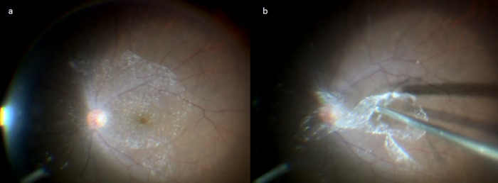
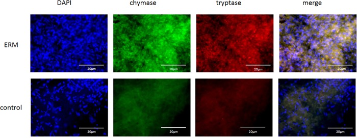
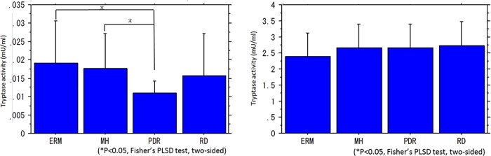
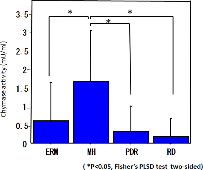
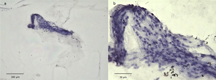
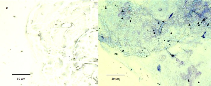
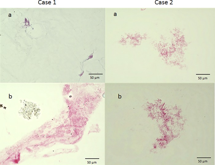
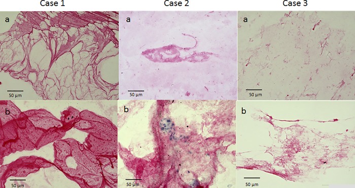
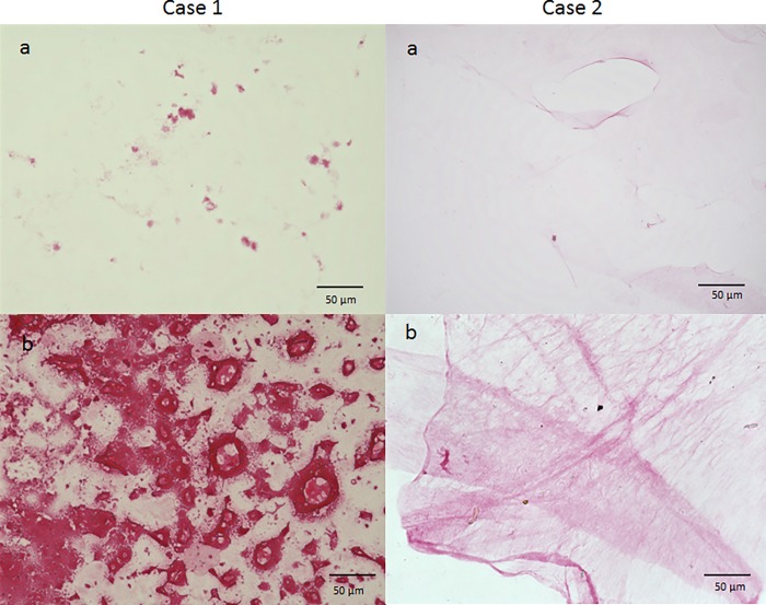
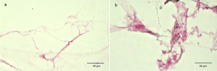
We previously reported on the elevated intravitreal activities of tryptase and chymase in association with idiopathic epiretinal membrane (ERM) and idiopathic macular hole (MH). In this present study, we investigated the potential intraocular production of these serine proteases, and measured and compared tryptase and chymase activities in the vitreous body and serum in ERM, MH, proliferative diabetic retinopathy (PDR), and rhegmatogenous retinal detachment (RRD) patients. In addition, nuclear staining with hematoxylin and eosin (H&E) and mast-cell staining with toluidine blue were performed on samples of the vitreous core and bursa premacularis (BPM) of MH. We also performed immunostaining on the above two regions of vitreous samples for MH with anti-tryptase antibody, anti-chymase antibody, anti-podoplanin antibody, anti-lymphatic vessel endothelial hyaluronan receptor 1 (LYVE-1) antibody, and anti-fibroblast antibody. Moreover, we performed immunostaining with anti-tryptase antibody and anti-chymase antibody on ERMs collected intraoperatively. Tryptase activity in the vitreous body was significantly higher in ERM and MH than in PDR. However, no significant differences were observed in the tryptase activity in the serum among these four diseases. Chymase activity in the vitreous body was significantly higher in MH than in the other three diseases, yet chymase activity in the serum was below detection limit in any of the diseases. Nuclear staining with H&E revealed an abundance of nuclei in the BPM region, but few in the surrounding area. Mast-cell staining with toluidine blue revealed that the BPM showed metachromatic staining. In immunostaining with anti-fibroblasts antibody, anti-tryptase antibody, anti-chymase antibody, anti-podoplanin antibody, and anti-LYVE-1 antibody, the BPM stained more strongly than the vitreous core. Tryptase and chymase-positive cells were also observed in ERM. These findings revealed that the presence of mast cells in the BPM potentially represent the source of these serine proteases. Moreover, the BPM, as a lymphatic tissue, may play an important role in the pathogenesis of macular disease.
我们之前报道过特发性视网膜内膜(ERM)和特发性黄斑裂孔(MH)与组织蛋白酶和糜蛋白酶的眼内高活性有关。在本研究中,我们研究了这些丝氨酸蛋白酶的潜在眼内产生,并测量和比较了 ERM、MH、增生性糖尿病性视网膜病变(PDR)和孔源性视网膜脱离(RRD)患者玻璃体和血清中的糜蛋白酶和组织蛋白酶活性。此外,对 MH 玻璃体核心和黄斑前膜(BPM)样本进行了苏木精和伊红(H&E)核染色和甲苯胺蓝肥大细胞染色。我们还对 MH 的上述两个玻璃体样本区域进行了抗组织蛋白酶抗体、抗糜蛋白酶抗体、抗 podoplanin 抗体、抗淋巴管内皮透明质酸受体 1(LYVE-1)抗体和抗成纤维细胞抗体的免疫染色。此外,我们对术中采集的 ERM 进行了抗组织蛋白酶抗体和抗糜蛋白酶抗体的免疫染色。玻璃体中的组织蛋白酶活性在 ERM 和 MH 中明显高于 PDR。然而,在这四种疾病的血清中,组织蛋白酶活性没有显著差异。玻璃体中的糜蛋白酶活性在 MH 中明显高于其他三种疾病,而在任何一种疾病的血清中,糜蛋白酶活性均低于检测限。H&E 核染色显示 BPM 区域有大量核,但周围区域很少。甲苯胺蓝肥大细胞染色显示 BPM 呈现异染性染色。在抗成纤维细胞抗体、抗组织蛋白酶抗体、抗糜蛋白酶抗体、抗 podoplanin 抗体和抗 LYVE-1 抗体的免疫染色中,BPM 比玻璃体核心染色更强。在 ERM 中也观察到组织蛋白酶和糜蛋白酶阳性细胞。这些发现表明,BPM 中的肥大细胞可能是这些丝氨酸蛋白酶的来源。此外,BPM 作为一种淋巴组织,可能在黄斑疾病的发病机制中发挥重要作用。